Archives

Herbs

Horehound Herb

A common and desirable benefit of the horehound herb is its effectiveness as an expectorant to aid the discharge of congestive materials from the lungs. This is but one of the many ways that horehound has been used to support good health throughout history. Since its first documented use in Roman times, many people have found it easy to gain health benefits from horehound since nearly every part of the plant is usable and there is a variety of preparation methods.

General Information About Horehound

White horehound is a bitter herb from the mint family that grows like a weed in many areas of the world. Horehound is an aromatic green plant, which produces many branching, square stems that often outgrow other field vegetation to be nearly two feet tall. Pairs of one-inch long leaves grow out of the stems in opposite directions. Between June and August, clusters of densely packed, small, white flowers bloom around the stems where the pairs of leaves attach. The flowers become seed-containing burrs, which have tiny barbs, allowing the seeds attach to animals, clothing, and machinery.
White Horehound.
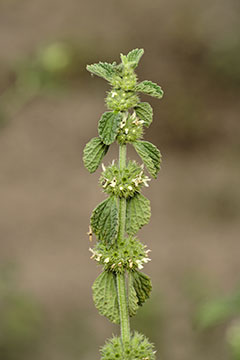
The hoary or silvery-colored hairs on its stem and give it a fuzzy or cloudy appearance are likely the reason for its English name, horehound. In old English, “har” and “hoary” mean grey or grey-haired. Its Latin name is Marrubium vulgare, which may have been derived from the name of an ancient Roman town, “Mariaurbs,” or from the name of one of the bitter herbs, “marrob,” used by the Jews during Passover. (Water horehound, called bugleweed, and black horehound, which has a strong, unpleasant taste and odor, are related to white horehound but the information here applies only to white horehound.)

Some suggest that the Egyptian god Horus could be a reason for its name. Priests in ancient Egypt may have called horehound the seed of Horus and used it in an antidote formula for poison, as did Caesar in the Roman era. Legend indicates that horehound aided priests during rituals in Egypt. Ancient lore gives horehound a power to break magic spells.

Early Physicians Recommend Horehound

Recorded mention of horehound began in the first century in ancient Rome. In his manual of medicine, Roman medical writer A. Cornelius Celsus, described antiseptic uses as well as treatments for respiratory ailments using horehound juice. In his book, “On Agriculture,” first century agriculturist Lucius Columella detailed how to use of horehound for various farm animal ailments such as ulcers, worms, and scabs. In the second century, the noted physician Galen also recommended using horehound to relieve coughing and to support respiratory health.

In his 1597 book on the history of plants and their uses, the respected British herbalist John Gerard recommended horehound as an antidote to poison and a syrup of horehound for those with respiratory problems. English physician Nicholas Culpeper echoed Gerard’s promotion of horehound in his 1652 book for physicians, stating, “There is a syrup made of this plant which I would recommend as an excellent help to evacuate tough phlegm and cold rheum from the lungs of aged persons, especially those who are asthmatic and short winded.”

The Spread of Horehound

Being a hardy perennial that grows well in dry soil, this highly recommended plant did not need help to spread beyond the Mediterranean region. Over the centuries, horehound flourished in all of Europe, South Africa, India, and other parts of Asia. It is now widely distributed in North and South America and Australia, although horehound is not native to these continents. Aiding the spread of horehound even more is the herb’s bitterness, which resists animal grazing. Horehound can be invasive, as in Australia where it is considered a bothersome weed.

Horehound’s reputation spread as well. Taking horehound for a cough or a cold was one of the natural remedies used by the early settlers of Australia. 18th century American physicians who recommended herbs touted its value for those with respiratory ailments and for menstrual problems. In the 1800’s in North America, horehound was used for those with hysteria and lung problems.

Traditional Uses of Horehound

Native American tribes have had many uses for horehound. Records show that ten tribes used it to treat various respiratory ailments, including two tribes which had a specific mixture for children’s colds. Some tribes used also horehound as a kidney flush, as a skin ointment, and as an antidiarrheal. These traditional Native American remedies were prepared from the leaves and flowers of the horehound and sometimes the root or the whole plant. Horehound was taken in the form of teas, extracts, and syrups for internal use and salves or poultices for external use. The Navajo tribe found additional value in horehound for stomach aches, influenza, infection, and as a gynecological aid before and after childbirth.

More recently, in 1956, Chopra, Nayar, and Chopra authored a book on medicinal plants in India. This book is cited a 2007 antibacterial research article (referred to below). The article acknowledges the book’s claims that, in addition to its value for the respiratory system, horehound “possesses tonic, aromatic, stimulant, diaphoretic [increase perspiration] and diuretic [increase urine flow] properties. … It was formerly much esteemed in various uterine, visceral, and hepatic [liver] affections….”

Other beneficial results have been claimed for the use of horehound. These claims include: relieving pain, promoting good digestion, reducing bloating, improving the appetite, stimulating bile flow, lowering blood pressure, vasorelaxant, reducing spasms (antispasmodic), killing intestinal parasites, easing morning sickness, relieving nausea or vomiting, and more. Of course, relief from these conditions is not guaranteed and a qualified healthcare practitioner should evaluate any serious symptom.

The support for these favorable effects has been largely anecdotal, and based upon tradition, scientific theory, or the reputation of horehound as an effective folk remedy. These reasons were sufficient to encourage the use of horehound well into the 20th century. In the industrial age, its reputation led to the production of extracts, teas, cough drops, and cough syrups containing horehound per some folk recipes.

Horehound’s Effectiveness Lacks Clinical Proof

Limited Clinical Evidence

Early in 21st century, however, few Americans seem to know anything about horehound or its history of supporting health. Despite recommendations and assertions of beneficial usage over many centuries, the use of horehound has surprisingly little support in clinical research and medical studies. Double blind human trials are yet needed to confirm or deny the valuable benefits that have been claimed for horehound. Neither has modern research made definitive findings concerning the toxicity, side effects, or proper dosage of horehound.

FDA Ruling on Horehound

Aiding horehound’s fall into relative obscurity was the 1989 FDA ruling on the use of non-prescription cough and cold medicines. The FDA approved just one of about 20 expectorant ingredients and did not find the others, including horehound, to be useful. According to this ruling, all non-prescription products containing the ingredients deemed ineffective must be taken off the market. However, cough suppressant products made outside the US which contain horehound, such as Ricola, continue to be sold in the US.

German Ruling on Horehound

In 1990, one year after the FDA’s ruling in the US, Germany’s Commission E listed loss of appetite and dyspepsia (indigestion) as acceptable uses for horehound. The 24 research scientists appointed to served on Commission E were to determine safe and effective herbal medicines; only approved herbs would be legal to sell in Germany. In identifying active components in horehound, the committee wrote that “[marrubiin] acts as a gastric juice stimulant and marrubinic acid acts as a choleretic [bile stimulant].” They found no known side effects, contraindications, or drug interactions. Notably, Commission E approved horehound to support gastrointestinal health, but they did not stand in opposition to the FDA ruling by approving horehound to support respiratory health, which they identified as unproven folk medicine.

Clinical Research Has Been Done on Horehound

Because clinical proof is now required and because of the FDA ruling, past claims made for horehound’s effectiveness are called into question. Yet, Marrubium vulgare (white horehound) has been the subject of some clinical research and, in light of the claims made for horehound, it is important to review these studies. While they are certainly not proof, in fact the evidence is said to be very weak, many publicly available online studies seem to offer support for horehound’s tradition of beneficial usage.

A health site quite reserved in its promotion of horehound, noted that “there is promising early evidence favoring the use of white horehound as a hypoglycemic agent for diabetes mellitus, and as a non-opioid pain reliever.” The following review of clinical findings on horehound will start with those two possible benefits.

Clinical Research on Horehound or its Ingredients

Hyperglycemia (Diabetes Mellitus) and Horehound

First, two projects show that horehound maybe effective against diabetes. From a 1992 study testing the hypoglycemic effect of 12 antidiabetic plants used in Mexico, “eight of the studied plants decreased significantly the hyperglycemia in rabbits as compared with control test (water). The strongest effect was yielded by Guaiacum coulteri, followed by Marrubium vulgare.”

In 2001, the hypoglycemic effect of five Brazilian medicinal plants was tested in rats. Four of the plants studied, including Marrubium vulgare, “significantly lowered the blood glucose. These results suggest that these four medicinal plants could be an adjuvant [helping] agent in the treatment of diabetes mellitus.”

Pain Relief and Horehound

Researchers in Brazil analyzed the “antinociceptive [pain-relieving] profile of marrubiin, the main constituent of [Marrubium vulgare] … in mice.

The results showed that marrubiin exhibits potent and dose-related antinociceptive effects. … the results suggest that marrubiin, like hydroalcoholic extract of M. vulgare, does not interact with opioid systems.”

Dried Horehound Herb.

In 2005, another clinical study in Brazil sought “to obtain more active compounds” of “marrubiin, a furane labdane diterpene, which is the main analgesic compound present in Marrubium vulgare.”

The scientists successfully formed “marrubiinic acid and two esterified derivatives … Marrubiinic acid showed better activity and excellent yield, and its analgesic effect was confirmed in other experimental models of pain in mice, suggesting its possible use as a model to obtain new and potent analgesic agents.

Blood Pressure and Horehound

Research in 2001, investigated “the hypotensive [BP lowering] effects of the water extract of Marrubium vulgare L. … in rats.” The horehound “extract lowered the systolic blood pressure … [by inhibiting] the contractile responses of rat aorta to noradrenaline and to KCl. … Marrubium displayed vascular relaxant activity.” This effective action was not blocked by the “NO synthase inhibitor N-nitro-L-arginine.”

Additional research was based on this significant hypotensive finding. A 2004 study added “that, in addition to its antihypertensive effect, Marrubium water extract improved the impaired endothelial [thin layer of cells lining blood vessels] function.” Because “crude extracts of the aerial parts of Marrubium vulgare show a potent in vitro inhibition of KCl-induced contraction of rat aorta, a group scientists from Belgium and Morocco sought to determine “pharmacological information about these components” of horehound. Their work, published in 2003, found “marrubenol [a diterpene alcohol] and marrubiin as the most active compounds.”

In 2004, researchers in Belgium, knowing that “Marrubenol inhibits contraction of rat arteries by blocking L-type calcium (Ca2+) channels in smooth muscle cells,” went on to investigate “its interaction with binding sites for calcium antagonists.” They found that “as marrubenol inhibited the contraction evoked by KCl depolarization of intestinal smooth muscle … interaction with the phenylalkylamine binding site seems to account for the inhibition of L-type Ca2+ channels by marrubenol.”

Antibacterial Activity and Horehound

White and black horehound were among the 5 of 168 crude, unfractionated extracts from Italian medicinal plants that tested well against “methicillin-resistant Staphylococcus aureus, a common cause of skin and soft tissue infection (SSTI).” In their 2008 article, researchers “identified a significant correlation in anti-biofilm activity with medicinal plants used for SSTI. They called it a step “towards the development of new anti-biofilm drugs … controlling the effects of pathogenic bacteria without strong selection for drug resistance.”

In India, in 2007, “the antibacterial activity of the methanolic extract of Marrubium vulgare whole plant was tested” “against selected strains … and exhibited moderate to significant antibacterial activity against five out of six tested bacterial organisms.” The researchers “inferred that the methanolic extract of M. vulgare whole plant had in-vitro antibacterial.”

References to Other Horehound Effects

Two of the above studies used to crude or simple water extracts, which a tea prepared at home could contain. One acknowledged that “chemically, [Marrubium vulgare] is best known for its … marrubiin, which has potent antinocicetpive [pain reducing] and expectorant effects.” Additionally, the 2008 article on Italian plants stated that “previous studies have demonstrated that white horehound extracts exhibit antispasmodic [and] antioxidant … properties.”

In 2001, a researcher studied marrubiin production because “horehound … has been used for centuries to relieve respiratory and bronchial ailments, and a compound known as marrubiin has been implicated as the active constituent.” The 2001 study in Brazil noted that “marrubiin … also is stated to stimulate secretions of the bronchial mucosa and to possess anti-arrhythmic properties.”

Together with the clinical summaries, such statements show that a large number of the traditional claims for the effectiveness of horehound are not dismissed by scientists, but are clearly acknowledged as a reason for their further research. Yet, it is also clear that much more research is needed to prove horehound effective by FDA standards.

How White Horehound Can Be Used

Preparation of Horehound

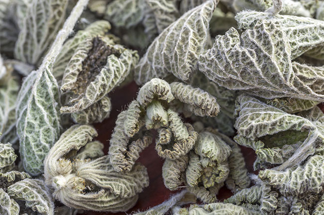
The entire plant can be used medicinally. Horehound is available fresh, dried, powdered, in capsules, as an extract, or as a pressed juice. When harvesting horehound, cut the plant when the buds of the flower appear. Immediately chop the horehound and then seal it in jars as soon as it has dried. Horehound can be made into candies, syrups, teas, and used as a flavoring.

Simple Recipes for Horehound

For these recipes, adjust amounts to strengthen to taste. Since horehound is quite bitter, most will add sweetener to taste.

Water Extracts

  • Tea: Pour boiling water on dried or bruised fresh leaves, one ounce of herb to a pint of water.
  • Infusion: Pour two cups of boiling water onto 1-2 ounces of dried horehound, cover and allow to steep for 10-15 minutes then strain.

Alcohol Extracts (Tinctures)

In a glass jar, soak fresh leaves in alcohol. Store in a dark place and shake the mixture several times daily. After about two weeks, strain the liquid with cheesecloth and store it in a tightly-sealed, dark glass bottle, preferably one with an eye-dropper.

  • For an extract, dilute fresh horehound with 20% ethanol in a 1:1 ratio.
  • For a tincture, dilute fresh horehound with Vodka in a 1:5 ratio.

Sweets

  • Snack: Mix chopped, fresh horehound with a little honey to chew and swallow.
  • Candy: Add sugar to an infusion of the leaves and boil it thickens. Pour it into a pan and cut into squares after it cools.
  • Syrup: Begin with a double-strength infusion using the fresh herb and add 24 ounces of sweetener – 12 ounces each of honey and brown sugar – for each 2.5 cups of the horehound infusion. Heat and stir the mixture as it thickens. After the mixture cools, refrigerate in glass bottles.

Daily Usage Suggestions

  • Syrup: one teaspoonful three times a day or 2-4ml.
  • Dried herb: 1-2 grams or by infusion.
  • Fresh leaves: 4.5 grams.
  • Juice: 30 to 60ml.
  • Ethanol Extract: 1-2ml
  • Tincture: 3-6ml or 10–12 drops in water up to three times a day.
  • Capsule: 1 containing 750ml horehound

Other Horehound Uses

An infusion is sometimes used externally as a wash, or a salve of horehound salve can be prepared, to disinfect wounds or for minor skin irritations. A cold infusion of white horehound acts is said to stimulate the bile flow of bile, while a warm infusion could aid sweating or possible break fevers.

Horehound Price Approximations

  • Online retailers sell horehound in various forms. Prices vary with retailer.
  • Liquid Extract: 7$ per ounce.
  • Cut tea: $1 per ounce, cheaper in bulk.
  • Cut and Sifted: $8 – $12 for 1 pound and $12 – $25 for organic.
  • Tea bags: 4$ for 25 bags.
  • Candy: $15 for 12 ounce tin or $3 per pound in bulk.
  • Capsules: $5 for 60 capsules containing 750mg horehound leaves.

Information on the Action of Horehound

Components

Horehound’s main active compound is the diterpene lactone marrubiin, as identified in the research summarized above. Many of its other components include:

  • A volatile oil with camphene and limonene
  • Diterpene alcohols such as marrubenol and marrubiol
  • Sterols
  • Saponins
  • Tannins
  • Mucilage
  • Alkaloids such as Betonicine and Choline
  • Flavonoids such as luteolin, quercetin, and their glycosides
  • Vitamin C
  • Potassium

Side Effects and Warnings

Horehound has a tradition safe usage; its effectiveness is the main reason for concern. If it works as a digestive aid, horehound use may upset people with ulcers or stomach problems. Also, excessive use of horehound may increase the risk of abnormal heart rhythms. Additionally, in contrast to some traditional uses, some recommend not using horehound during pregnancy or breastfeeding, and should not be used with infants.

Interactions

Because of its possible effectiveness, it is best to err on the side of caution and consult the prescribing doctor before using horehound. Below is a partial list of possible interactions:

  • Because horehound may work to lower blood pressure, those taking blood pressure medication should use caution.
  • Similarly, horehound’s possible effectiveness as a diuretic and in lowering blood sugar, caution should be used when taking a diuretic medication such as water pills or a medication that affects blood sugar.
  • Because horehound may work as an expectorant, it may change or increase the effect of cold medications.
  • Similarly, horehound’s possible effectiveness may increase the effect of laxitive products or cholesterol-lowering medications.
  • Because horehound contains glycosides and estrogen-like chemicals, those taking heart medications or hormone therapy should use caution as well.
  • Additionally, those who take supplements or other herbs to address a condition that horehound may affect should use caution and consult their health practitioner.

References for the article

Clinical Research References

Hyperglycemia (Diabetes Mellitus) and Horehound
Roman Ramos R, Alarcon-Aguilar F, Lara-Lemus A, et al.
Hypoglycemic effect of plants used in Mexico as antidiabetics.
Arch Med Res . 1992;23:59–64

Novaes AP, Rossi C, Poffo C, Pretti Júnior E, Oliveira AE, Schlemper V, Niero R, Cechinel-Filho V, Bürger C.
Preliminary evaluation of the hypoglycemic effect of some Brazilian medicinal plants.
Therapie . 2001;56:427–30

Pain Relief and Horehound
De Jesus RA, Cechinel-Filho V, Oliveira AE, Schlemper V.
Analysis of the antinociceptive properties of marrubiin isolated from Marrubium vulgare.
Phytomedicine. 2000 Apr;7(2):111-5.

Meyre-Silva C, Yunes RA, Schlemper V, Campos-Buzzi F, Cechinel-Filho V.
Analgesic potential of marrubiin derivatives, a bioactive diterpene present in Marrubium vulgare (Lamiaceae).
Farmaco. 2005 Apr;60(4):321-6.

Blood Pressure and Horehound

El Bardai S, Lyoussi B, Wibo M, et al.
Pharmacological evidence of hypotensive activity of Marrubium vulgare and Foeniculum vulgare in spontaneously hypertensive rat. Clin Exp Hypertens . 2001 May;23(4):329-43.

Sanae El Bardaia, b, Marie-Christine Hamaidea, Badiaa Lyoussib, Joëlle Quetin-Leclercqc, Nicole Morela and Maurice Wibo
Marrubenol interacts with the phenylalkylamine binding site of the L-type calcium channel

Antibacterial Activity and Horehound

Cassandra L. Quave, Lisa R.W. Plano, Traci Pantuso, Bradley C. Bennett
Effects of extracts from Italian medicinal plants on planktonic growth, biofilm formation and adherence of methicillin-resistant Staphylococcus aureus
Journal of Ethnopharmacology: Accepted 8 May, 2008

It also had sources for antispasmodic (Schlemper et al., 1996), antioxidant (Berrougui et al., 2006; Matkowski and Piotrowska, 2006; Weel et al., 1999)

Mubashir H. Masoodi1*, Bahar Ahmed2, Iqbal M. Zargar1, Saroor A. Khan2, Shamshir Khan2
and Singh P.1
Antibacterial activity of whole plant extract of Marrubium vulgare
African Journal of Biotechnology Vol. 7 (2), pp. 086-087, 18 January, 2008

Other Benefits Acknowledged in Research

Researcher wins accolades.

Interactions

Horehound Interactions

Conditions

Plantar Fasciitis

The plantar fascia is a sheet of very tough material running the length of the bottom of your foot. Basically a long and flat but thick sheet of connective tissue, it attaches your heel to each of your toes. Tissues that connect bones to other bones are called ligaments, so this may also be referred to as the plantar fascia ligament.

Like other connective tissue, it is constructed primarily of a protein called collagen, in this case type I collagen. Other elements of the plantar fascia include: elastin; fibroblasts, which secrete collagen; glycosaminoglycans or GAGs, which are carbohydrates that aid in resilience; water, nerves and blood vessels.

Plantar Fasciitis.

About Plantar Fasciitis

The condition called plantar fasciitis occurs when small tears or ruptures occur in the plantar fascia, causing pain especially on the bottom of the heel. The plantar fascia can also be termed the plantar aponeurosis. When the condition goes beyond inflammation and the damage becomes long-lasting, it is sometimes called plantar fasciosis. A more serious but much less common injury is plantar fascia rupture, which is a serious tear to the entire fascia rather than just micro injuries.

Something else that can damage the plantar fascia are heel spurs. These are sharp pieces of bone formed where the plantar fascia has been pulling away from where it is attached to the heel. Bony tissue is laid down at the points of separation. These can then dig into the surrounding softer tissues.

Symptoms

The pain from plantar fasciitis usually develops gradually and in one foot rather than both. It is felt right in the middle of the bottom of the heel where the plantar fascia has its base of attachment to the heel bone (calcaneus). Pain upon awakening is typical, with eventual lessening with activity. Often standing for a long time or abruptly arising from a seated position can worsen the pain.Foot Problems Plantar

Causes

The plantar fascia does more than just attach the heel bone to the toe bones. It also supports the other bones of the foot and has a biomechanical function. It can expand and contract with striding, acting as a springboard to help impel motion and helping to absorb the shock of impact with each footfall. It tensions the bottom of the foot between the heel and the toes.

It is the structure of collagen itself that enables much of this biomechanical function. The collagen is laid down in an ordered way and the strands of collagen I are arranged in a braided or rope-like manner which allows for strength and elasticity. If conditions arise which place undue stress on the plantar fascia, collagen strands can break and begin to unravel.

This results in small tears or ruptures within the plantar fascia. Fibroblasts produce new collagen to repair the damage, but it is collagen III which is not as strong or flexible as collagen I. Eventually the collagen III is replaced by collagen I, but as is typical with connective tissue, healing is slow to occur.

It used to be thought that the pain of plantar fasciitis was caused by the presence of heel spurs, since these were frequently found during diagnostic imaging. However, it has been found that people can have heel spurs without the painful symptoms, which calls this into question. If usual treatments for treating plantar fasciitis don’t bring relief, pain from bone spurs present should be investigated.

Risk Factors

Conditions which can cause tears or ruptures to occur in the plantar fascia are:

  • Wearing ill-fitting or worn-out shoes.
  • Being overweight.
  • Having abnormalities in foot arch structure such as very high arches or low arches.
  • Having an abnormal gait, especially where the feet roll inward while walking (pronation, or having flat feet).
  • Exercise that stresses the heels of the feet, such as running and some types of dancing.
  • Needing to stand or walk on hard surfaces for long periods of time, such as in nursing, teaching, waitressing, military personnel.
  • Being over 40.
  • Being female.
  • Having arthritis, which can cause inflammation to tendons and ligaments.
  • Having diabetes.
  • Being pregnant.

Prevention Tips

Wear shoes that fit your feet and provide good arch support. Take care to replace athletic shoes before the cushioning wears out. If you run, shoes are good for about 500 miles. Avoid shoes with high heels, since these place stress on the heel and Achilles tendon, which in turn affects the plantar fascia. The Achilles tendon attaches the base of the heel to the calf muscles.

Devices which cushion and support the heel. Routine use of arch supports, orthotic devices and cushioning insoles can help prevent plantar fascia injury.

Body weight can be a factor as well so it is important to maintain a healthy body weight.

Pregnancy

With the added weight gain during pregnancy and the presence of hormones which cause softening and relaxing of connective tissue, stress is added to the plantar fascia. Care should be taken during this time to not stress out the ligament.

Sudden Changes in Exercise Routines

Before making changes to more rigorous exercises or activities such as in running longer distances, adding extra hours of practice to field sports, or escalating to more demanding routines, consideration should be given to the possibility of injuring the plantar fascia.

Stretching exercises before being active loosens the ligament and helps prevent damage, as does a gradual increase in intensity and duration of activities rather than a sudden onset. As a person ages and tendons and ligaments become more inflexible in general, care should also be taken to stretch before activity.

Correcting Inborn Gait Problems

Working with a podiatrist and/or an orthotic specialist to analyze gait irregularities and then to produce devices to correct them can lessen the likelihood of plantar fascia injury over time.
Foot Problems Plantar
Sometimes, and especially for athletes, plantar fascia taping is recommended. Taping has to be carefully done using a special athletic tape and with the feet dry and clean. It is usually applied before activity that might stress the plantar fascia or at the beginning of a day to give the feet support in general. Tape should not be left continuously in place. It is often used in combination with orthotic supports.

Test & Diagnosis Considerations

The diagnostic heel pain which is worst upon getting out of bed is caused by the tightening of the plantar fascia during sleep. When activity is begun for the day, the ligament doesn’t move well and pain is the result. As movement warms the plantar fascia, pain often decreases. The pain is variously described as stabbing, tearing, or burning.

A physician will often press on the midheel area where the ligament joins with the heel bone to affirm the source of the pain. The foot is examined to check for swellings, tender areas and inflammation. X-rays or MRIs may be taken to rule out bone stress fractures or pinched nerves.

Pain in other areas of the foot such as the ball of the foot should not be confused with plantar fasciitis. Pain in the ball of the foot is usually associated with metatarsalgia, which is inflammation of the tissues which accompany the bones of the arch of the foot (metatarsals). Pain along the back of heel up toward the calf muscle is usually associated with Achilles tendon problems.

Treatment Options

It is important to begin treatments as soon as possible after symptoms are noted to prevent serious lasting damage to the plantar fascia. Symptoms can usually be alleviated by conservative non-surgical methods, but it can take a lengthy period of time to reverse the damage.

One of the possible consequences of not seeking prompt treatment is developing gait changes that can affect other parts of the body while attempting to favor the injured foot. Ankle, knee, hip and back problems can follow untreated plantar fasciitis.

Reducing Inflammation

Immediate icing of the painful area is helpful to reduce pain and swelling. Use something that can conform to body contours such refreezable gel packs or ice bags. Apply the ice for 20 to 30 minutes until numbness sets in; then remove for 20-30 minutes before reapplying. A combination of applying cold while stretching the plantar fascia is of benefit. Freeze water in paper cups or use a can of frozen juice. Roll one of these about under the affected foot, massaging and flexing the bottom of the foot while numbing with coldness.

Taking non-steroidal anti-inflammatory medications such as ibuprofen, aspirin, and naproxen help relieve symptoms and lessen pain. They do not affect a cure, but they usually allow a person to be more comfortable.

Stretching & Flexibility

Increasing the flexibility of the plantar fascia can help too. There are a series of foot stretches and exercises that can be performed as well as calf exercises. One exercise is to sit down, extend your leg in front of you with the knee bent and grasp the toes in one hand. Gently pull them back toward the top of the foot until you feel a pull in the arch area. This lengthens and stretches the ligament.

A towel stretch accomplishes much the same thing. Loop a rolled towel under the ball of your foot and grasp the ends of the towel in your hands. Gently pull the towel toward your body, keeping the knee straight. Hold for 15-30 seconds and repeat three or four times.

The towel curl involves sitting down, putting a towel on the floor under the foot and using the toes to gather the towel in folds, push the folds behind the foot. Then work in reverse, using the toes to push the towel forward ahead of the foot. Alternatively, you can also use your toes to “walk” your foot forward across a carpeted surface and then back toward you again. A related exercise is placing some marbles and a cup on the floor. Seated, try picking the marbles up with your toes and placing the marbles in the cup.

Additional Stretching

A simple plantar fascia/calf stretch uses stairs which have a banister or railing. Stand on one stair with the toes and ball of your feet on the tread, allowing the heels to suspend over the lower stair. Hold on to the railing or banister while doing this. Then relax your calf muscles and allow the heels to droop downward. You should feel a pull all along the bottom of your foot, through the back of your leg and to the knee. After about 30 seconds, tighten the calf muscle slightly to raise the heels again. Repeat this several times.

Another stretch is for the calf and Achilles tendon. Facing a wall, brace your hands against it, arms extended at about chest height. Step back with the damaged foot and place the foot flat on the floor. Step forward with the other foot and bend it gradually so you lean toward the wall. When you feel the stretch in your calf down toward your heel, hold for 15 to 30 seconds, then relax. Repeat this several times.

Working the Arch of the Foot

To exercise the area in the arch of the foot, sit down and put a tennis ball or rolling pin on the floor and roll it around using the area in the arch of the foot. If you can balance well, you can try doing this exercise while standing.

All exercises can be repeated several times a day for best results in strengthening the fascia and making it more flexible.

Massage is sometimes helpful. Often people undergo physical therapy to aid in recovery. A physical therapist can work with each individual to provide exercises that work for his or her set of particular circumstances. Acupuncture has also been used to treat persistent pain.

Losing Weight

Weight loss can often alleviate symptoms. Use of orthothic supports which fit inside the shoe can help cushion the heel, distribute weight better and encourage proper gait while the foot is healing. Both custom-made ones for your individual foot and off-the-shelf models work. Favor your feet by not going barefoot, even when you get up for brief periods at night. Slip on a pair of shoes which will provide arch support even for going short distances.

Resting the Foot

Another way of removing the stress, especially when severe pain is present, is to rest the foot, giving it a chance to recover from too much running, jumping, walking, dancing, or whatever behavior brought on the injury. For less severe pain, minimizing the activity for a while until pain abates is advisable. Also consider switching high-impact activities such as jogging and running for low-impact activities such as swimming or riding a stationary or regular bicycle for a while.

Taping of plantar fascia is sometimes done to help alleviate pain, although usually it is considered as primarily a preventative measure (which see).

A device known as a night splint can help lessen the pain upon awakening. The padded night splint fits along the bottom of your lower leg and the bottom of your foot, with straps around your leg and foot holding it in place. This support keeps the plantar fascia stretched while you sleep or rest.

Corticosteroid Use

If these conservative treatments do not work, injections of corticosteriods directly into the painful area may be considered. Steroids act to decrease inflammation and thus reduce pain. Injections do not always solve the problem and may worsen it by further weakening the connective tissue. Another method of providing corticosteriods to the injured area is by putting them on the skin surface and then using an electric stimulus to aid in absorption through the skin. This is called iontophoresis and is not a painful procedure.

Extracorporeal Shock Wave Therapy (ESWT)

Shock waves (sound waves) are used to bombard the injured area to stimulate it and hopefully hasten repair and recovery of the connective tissue. This is not a widely used treatment and it is still being assessed. It can cause numbness, tingling, swelling or pain over the area in which it has been used.

Surgery

The treatment of last resort is surgery. If the tissue has been so damaged that repair is unlikely, surgery to disconnect the ligament from the heel bone can be performed. Usually this is done mostly to lessen chronic severe pain when nothing else has worked. Understandably this procedure weakens the foot arch because the tension between toes and heel that the plantar fascia provides is no longer there. Side effects of the surgery can be infection, nerve injury, rupture of the plantar fascia and also no relief of pain.

After surgery support in the form of a brace or cast is needed for a varying length of time for proper healing of the tissue, with several weeks usually being needed before weight can be applied to the heel again. The usual recovery time to normal foot use is about three months.

Endoscopic surgery may also be used for a less invasive procedure. This is where small incisions are made to allow the passage of tools and a lighted camera. This procedure allows location and removal of the damaged part of the plantar fascia with less injury to surrounding tissue.

Coblation Surgery

A newer surgical technique than traditional surgery that has been used for treating plantar fasciitis is coblation surgery. The name is derived from “controlled ablation.” The technique is also called the Topaz procedure. It uses radiofrequency energy combined with a saline solution to remove tissue gently and with minimal destruction to adjacent structures.

If a heel spur seems to be causing pain by pressing into tender tissues near the bottom of the heel, steriod injections can be used here as well followed by surgery to remove the spur if injections do not relieve the pain.

Additional Resources

About.Com Treatment Options
Mayo Clinic, Plantar Faciitis Information
American Academy of Podiatric Sports Medicine

Conditions

Menstrual Cramps

Menstrual cramps experienced by a woman during her monthly menstrual period are pains that occur in the abdomen and pelvic area. To some extent, 50 percent of all women are affected by menstrual cramps and approximately 15 percent of these women have severe cramps. Ninety percent of girls still in their adolescence have cramps.

Menstrual cramps can be mild to severe and are different in every woman who has them. Mild cramps are short in duration and are sometimes only slightly noticeable. But some women suffer such severe cramps that it can interfere with their daily activities and may last for several days.

Menstrual Cycle.

Types of Menstrual Cramps

Menstrual cramps can be one of two types: primary or secondary. Primary cramps are the type of menstrual cramps that are most common. Women can begin to have them with the onset of a their first period or they can begin after several years. They are caused by no physical abnormality of any kind. The secondary type of menstrual cramps have an underlying physical basis like pelvic inflammatory disease (PID), fibroids in the uterus, and other conditions discussed later.
Menstrual Cramps

Alternative Names

The medical term for common menstrual cramps is secondary dysmenorrhea and is often just referred to as dysmenorrhea. When there is underlying cause of the cramps, it is referred to as secondary dysmenorrhea.

Symptoms

The pain from menstrual cramps starts in the lower abdomen and the pelvis and can extend to the lower back as well as the legs. For some women the pain may be only a dull ache while for others they are more painful.

Menstrual cramps usually begin before the period actually starts and will normally be the worst about 24 hours after they begin. They generally subside after one to two days. Other symptoms that may accompany cramps are:

  • Headache
  • Nausea, which can rarely be followed by vomiting
  • Constipation
  • Diarrhea (The prostaglandins cause the muscles of the intestinal tract to contract.)
  • More frequent urination.

Primary Dysmenorrhea

Primary dysmenorrhea happens when the endometrium, which is the inner lining of the uterus, sheds as the menstrual flow each month. During the month, this lining is built up in order to make the uterus ready for a possible pregnancy. When an egg is not fertilized during ovulation to cause a pregnancy, the inner lining is not needed. The levels of estrogen and progesterone in the woman’s body decrease, the uterine lining swells and is released from the body as menstrual flow. When this is completed, the lining is replaced.

During the break down of the uterine lining, compounds known as prostaglandins are discharged, causing the muscles in the uterus to contract. This contraction causes less blood and oxygen to flow into the endometrium which makes it break down and die. When the tissue is dead, the contractions continue in order to push the dead tissue through the cervix and away from the body via the vagina.

Levels of other substances known as leukotrienes increase during this time as well. These chemicals are connected to the body’s inflammatory responses and may also play a role in causing menstrual cramps.

Cramps caused by this monthly process can be particularly severe when pieces of bloody tissue and clots are pushed out through the cervix. Women with a narrow cervical canal may experience these severe cramps.

The severity of cramps can often depend on the level of the prostaglandin in the body. Women with a higher amount of prostaglandin will have cramps while a woman with normal to low levels may not experience menstrual cramps.

There are other issues that can add to the severity of menstrual cramps. Emotional stress has been shown to increase the discomfort felt from cramps, as well as a lack of exercise.

Menstrual cramps caused by primary dysmenorrhea will normally lessen as the woman ages or after giving birth. It is believed that the nerves inside the uterus degenerate as they age. In a pregnant woman, these nerves deteriorate completely in the later stages of the pregnancy and only some of them are restored after childbirth.

Secondary Dysmenorrhea

Secondary dysmenorrhea is caused by some condition usually connected to the reproductive system and usually develops later in life rather when an adolescent begins her menstrual periods. Some of these conditions are:

  • Endometriosis – This is a condition in which the tissue of the uterine lining affixes itself to other areas not in the uterus, causing lesions.
  • Uterine polyps – These are growths that are attached to the inner wall of the uterus and protrude into the cavity of the uterus. They are caused by too much growth of cells in the endometrium.
  • Pelvic inflammatory disease – This is an infection of the reproductive organs.
  • Uterine fibroids – These are growths in the uterus whose development may be stimulated by estrogen and progesterone.
  • Using an IUD for birth control.

Risk Factors

There are people who may be at a higher risk of primary menstrual cramps each month. This includes:

  • ·Women who are under the age of 20
  • Women who experienced an onset of puberty at age 11 or younger
  • Having particularly heavy bleeding with their periods
  • Having never been pregnant nor gone through childbirth
  • Being significantly overweight or underweight.
  • Smoking. Smokers have a 50 percent higher chance than women who don’t smoke of suffering from menstrual cramps.
  • Women under high levels of stress.

Prevention Tips

There are ways that may work for some women to keep them from having menstrual cramps or at least reducing their severity. Many of these prevention tips are naturopathic solutions and can be used as treatments when menstrual cramps do occur.

  • Get regular aerobic exercise.
  • Talk to your doctor about taking birth control pills in order to stop the production of hormones that contribute to menstrual cramps.
  • Cut back on coffee, decaffeinated coffee, and black tea, especially in the days before the period begins.
  • Cut back on alcohol consumption.
  • Reduce the amount of meat and dairy food in the diet. They can aid in the production of the prostaglandins that help to cause cramps.
  • Cut back on salt before the period begins.
  • Take a vitamin supplement that contains B3, E, magnesium citrate, and calcium citrate.
  • Eat cold-water fish and/or take a fish oil supplement with Omega 3 and fatty acids. Doing this can inhibit the production of prostaglandins.
  • Practice certain yoga positions to aid the proper balance of the uterine function.
  • Avoid the use of intra-uterine devices (IUDs) as a method of birth control. These devices can increase menstrual cramps.
  • Use sanitary pads instead of tampons.

Test and Diagnosis Considerations

A woman can generally diagnose menstrual cramps herself and it is based on her own perception of the pain. In the case of primary dysmenorrhea, most women know what the signs and symptoms are. However, women who experience more than two painful periods, particularly if the pain is disabling, should schedule an appointment with their gynecologist between periods.

In these cases, the gynecologist will likely perform a pelvic exam to ensure that there are no other causes for the pain.

For older women it is possible that the pain they are feeling is secondary dysmenorrhea and may be due to an underlying issue. Besides a pelvic exam, the doctor may perform an ultrasound to look for unusual growths or cysts on any of the reproductive organs.

If the cause of the cramps is undetermined after an ultrasound, the doctor may also insert a scope through a small cut below the navel in order to perform a more thorough examination of the region. This is called a laparoscopy.

A D&C, which is discussed in the section on Surgical Treatments is sometimes used to diagnose uterine conditions.

Treatment Options

Treatment for menstrual cramps can vary from woman to woman and may require some trial and error before finding the one that is right. Some of the prevention options mentioned previously work for many women like regular exercise, adequate rest and sleep, and yoga exercises. Heating pads and soaking in a hot bath have also been known to help.

There are various pharmaceutical treatments as well as herbal and home remedies and very rarely, surgical treatments that help relieve menstrual cramps. For each treatment, the level of efficacy may be different in every woman.

Pharmaceutical

There are several non-prescription drugs that can help control the pain of menstrual cramps, and in some cases, even prevent them. Aspirin and acetaminophen are enough for some women who have mild cramps. There are over-the-counter drugs made specifically to treat cramps that contain acetaminophen plus a diuretic.
Menstrual Cramps
Non-steroidal anti-inflammatory drugs (NSAIDs) are the best non-prescription option to treat menstrual cramps. Drugs that fall into this category are ibuprofen, naproxen, and ketoprofen.

It is best to begin taking these before the onset of cramps so the pain does not get too difficult to control. This could mean taking the pills as early as one or two days before the periods is due to start.

The medication should be taken one to two days after the period begins. There are also prescription strength NSAIDS that are available for treatment of menstrual cramps.

Oral Contraceptives

A method often used to treat severe menstrual cramps is taking oral contraceptives. Birth control pills contain estrogen and progestin and prevent ovulation, thus decreasing the amount of prostaglandins that are produced. This can makes the cramps much less severe and has the added advantage of causing a lighter menstrual flow.

While the use of an IUD for birth control can make menstrual cramps worse, one treatment option entails the use of an IUD that releases progestin directly into the uterus. This treatment has been shown to reduce menstrual cramps by 50 percent.

Herbal and Home Remedies

Many of the preventative measures listed for menstrual cramps are also good home remedies for when the cramps do manifest. Those, plus other herbal and home remedies can help to ease the discomfort of painful menstrual cramps.

Some people turn to Acupressure, this practice is based on the principles used in acupuncture, but instead of using needles on the acupuncture points, light pressure is applied. This practice is often recommended by those who practice alternative medicine for the relief of menstrual cramps.

The proper acupuncture point is called Spleen 6 and is approximately the width of four fingers above the bony part of the inner ankle, next to the shinbone and towards the back part of the calf. Slowly increasing pressure should be applied to this point using either the thumb or middle finger. Hold this for 3 minutes.

  • Place a hot water bottle on your stomach. A damp towel heated in the microwave can also be used. This relaxes the muscles that cramp.
  • Abdominal cramping can be relieved through orgasm. The muscle contractions that occur during an orgasm make the blood and other fluids leave organs that are congested.
  • Drink two cups of peppermint or wintergreen tea a day. The mint leaves can be added to any type of tea. Also suck on mint candy on and off during the day.
  • Drinking hot liquids can increase blood flow and relax muscles that cramp. Drink any herbal tea or heat up lemonade. Don’t drink any hot liquids that contain caffeine. Caffeine increases nervous energy and can make menstrual cramps worse.
  • A hot bath with mineral salts can help relax muscles. Soak for at least 20 minutes.
  • Basil has a natural painkiller called caffeic acid and it can relieve the pain menstrual cramping. To make a basil tea, put two tablespoons of basil leaves in a pint of boiling water. Allow this to steep covered until it comes to room temperature. To ease cramps, drink ½ cup to 1 cup of the tea every hour.
  • Cinnamon has antispasmodic and anti-inflammatory properties. Sprinkle cinnamon in tea or on breakfast food.
  • Calcium is well known to decreasing pain from menstrual cramps. Taking magnesium supplements both before the period begins and during it can help the body more effectively absorb the calcium in food and calcium supplements.
  • Drink plenty of water. Dehydration can make menstrual cramps worse.
  • Getting plenty of rest can help to decrease cramps.

Home remedies have the added advantage of being affordable and easily accessible.

Surgical Procedures

Surgical procedures are not often recommended for the treatment of menstrual cramps. In cases when a woman consistently has uterine bleeding that is unusually heavy and painful, a doctor may perform a procedure called an endometrial ablation. In this procedure, the uterine lining is destroyed using a device that generates heat like a laser beam, electricity, freezing, microwave, or heat produced by radio frequency. After the procedure, scars cause the endometrium to heal and this significantly reduces uterine bleeding and the severe cramps that go along with it.

The average fee for an endometrial ablation is $3,516, which does not include the hospital and anesthesia fees.

Another procedure to reduce heavy bleeding and cramping that used to be done more commonly in the past than it is today is called dilation and curettage (D&C). During this procedure, the cervix is expanded and a surgical instrument is inserted and used to remove tissue from the endometrium. This instrument is either sharp to scrape out the tissue or it sucks the tissue out. A D&C has minimal side effects. The most common is cramping.

If performed in a physician’s office, the cost can be low as lows $400. Performed at a surgical facility it can be as high as $2,000.

Treatment

The treatment for this type of menstrual cramps may be different depending on what the underlying cause is. Once the condition is diagnosed, the doctor will recommend the proper treatment.

Conditions

Types of Edema

Edema is the accumulation of abnormal levels of fluid in the circulatory system and the tissues between the body’s cells (interstitial tissue). This condition often causes tissue underneath the skin to swell, and is common when lymphatic blockage occurs.
Edema Information
Swelling occurs in the blood vessels when an excessive increase in the volume of plasma fluid causes the vessels to dilate.

Eventually, blood vessel walls weaken as they dilate, causing lymph fluid to diffuse into surrounding tissue space.

This excess fluid gets trapped in the body’s tissue in a greater capacity than the lymphatic system can eliminate.

Generally, edema is physically unnoticeable until the interstitial fluid volume is approximately 30% above normal (normal fluid volume is 10%).

Many American’s suffer from edema without knowing what it is, and what causes it. Edema is also referred to as oedema, which was formally labeled dropsy or hydropsy.

Symptoms of Edema

Swelling of the Skin

  • Localized or peripheral: swelling is in one specific area. Mainly in one of the legs or arms.
  • Generalized: swelling is in particular regions of the body. Such as in the extremities, a portion or all of the face, abdomen, and the feet.
  • Organ-specific: swelling in a particular organ such as the brain (cerebral edema), the lungs (pulmonary edema) or the eye’s (periorbital edema).
  • Pitting Edema: a severe case of edema which exists if skin retains a dimple after being pressed for 10 to 20 seconds.

The skin may feel tight and stretched. Edema causes the skin to feel tight as a result of being stretched beyond its normal boundaries. In some cases, skin may either feel warm to the touch, or quite cool if there is a blockage in local circulation.

Additional Symptoms to Look For

You may also experience puffiness of the ankles. Boggy ankles are one of the most common signs of peripheral edema; which is characterized by generalized swelling in the extremities.

In addition to ankle swelling, you may also experience swelling of the face or eyes. This can cause the face to become red and swollen. Swelling of the eyes can occur in the cornea of the eye due to glaucoma, conjunctivitis, keratitis, or surgery. Periorbital edema causes puffiness around the eyes, and is most noticeable immediately after waking. Other issues to watch out for are:

  • Weight gain: due to increased fluid retention, and lack of elimination.
  • Increased localized heat radiating from skin’s surface: is commonly associated with a localized infection.
  • Hypertension: or an increase in blood pressure due to blood flow blockage resulting from an influx of fluid levels.
  • Stiff joints and aching body parts: are the body’s reactions to pain, signaling that a disorder has occurred.

Causes of Edema

There can be many causes of Edema, such as:

  • Physical inactivity
  • Genetic predisposition
  • Standing or sitting for long periods of time
  • Surgery resulting in localized inflammation or impaired organ functionalities
  • Burns
  • Heat
  • High Altitudes
  • Pregnancy
  • Certain medications
  • Excessive salt intake
  • Contraceptive Pill
  • Menopause
  • Poor diet
  • Kidney disease/damage
  • Heart failure
  • Chronic lung disease
  • Liver disease
  • Diabetes
  • Arthritis
  • Thyroid disease
  • Brain tumor
  • Head injury

Risk Factors for Edema

  1. Congestive heart failure – blood accumulation in the legs, ankles, or feet due to the hearts inability to pump efficiently.
  2. Cirrhosis – scarring of the liver that imbalances the secretion of hormones and chemicals regulating fluid levels in the body.
  3. Kidney disease – cause fluid and sodium retention in the legs and around the eyes.
  4. Nephrotic syndrome – kidney damage causing low protein levels in the blood to result in an over accumulation of fluid.
  5. Chronic venous insufficiency (CVI) – weakening of the veins and values in the legs, which causes them to lack the proper pumping abilities needed to push blood back to the heart.
  6. Deep vein thrombosis – a blood clot which forms in a vein, mainly in the thigh or lower leg.
  7. Lymphedema – the improper draining of lymph nodes and vessels due to genetic predisposition, or a medical condition such as cancer or an infection.

Tests and Diagnosis

In order to treat edema a doctor has to diagnose what is causing it. However, in cases showing signs of systemic edema (caused by a malfunction of one or more of the systems in the body) or pitting edema, it is wise not to wait to seek the advice of a medical practitioner.
Diagnosing Edema
The initial set of test done by a medical professional is a preliminary evaluation. First, a medical history profile is completed. Then, the practitioner performs a physical assessment, comparing patient’s arms and legs for symmetrical edema and other areas of the body for dependent edema. Last, the medical professional palpates the area affected by taking peripheral pulses, by notating areas of the body that are cold to the touch, and by performing a complete cardiac and respiratory assessment.

If edema is confirmed additional test are recommended, correlating to the area of the body in which the edema occurs.

Testing for Localized Edema

Localized Edema can be determined during a patient consultation, which includes health history questionnaires, and physical assessments carried out by a medical professional. Once localized edema is suspected the following test may be done to diagnose the exact cause:

  1. Impedance phlebography is a noninvasive test that uses electrical monitoring to measure blood flow in veins of the leg. Information from this test helps a doctor to detect deep vein thrombosis (blood clots or thrombophlebitis).
  2. D-dimer testing is a sensitive indicator for active deep vein thrombophlebitis.
  3. Cultures of blood or fluids that are available from the site of the lesion should be taken for patients showing symptoms of cellulitis or osteomyelitis.
  4. X-rays and CT scans should be taken of the involved area.
  5. Bone scans should be taken for patients thought to have osteomyelitis and bone fractures.
  6. Lymphangiography is used to determine if lymphedema exists.
  7. CT scan of the pelvis or abdomen is used when signs of malignant lymph nodes exist.
  8. Thyroid profile detects pretibial myxedema as a result of thyrotoxicosis.

Testing for Generalized Edema

Generalized Edema is systematic. Therefore, endocrine testing is one of the primary components used to make a diagnosis.

  1. Venogram is used to test edema in the upper extremities caused by increased venous pressure.
  2. Doppler or ultrasound study is also used to test venous obstruction of the upper extremities as well.
  3. Computed Tomography (CT) if thorastic outlet syndrome is present. Other options include Magnetic resonance imaging (MRI) or plain films.
  4. Chest X-ray (CXR) study and ECG are done to detect edema in the arms and legs.
  5. Liver function test are done if cirrhosis is suspected.
  6. Urinalysis and sediment evaluations are done if renal causes of edema are expected.

Organ Specific Edema Test

Organ Specific Edema test vary depending on the organ affected. However, general testing procedures still apply which include.

  1. Complete blood count (CBC)
  2. Urinalysis
  3. Chest films
  4. Electrocardiogram (ECG)
  5. Biochemical screening which include albumin, total protein, liver function tests, total cholesterol, and thyroid function tests

Testing for Pitting Edema

Press and hold the skin for 10-20 seconds. If the dimple impression remains when released then pitting edema may exists. If pitting edema exists for more than 3 months, low serum protein levels may be an underlying factor.

Prevention Tips

At home treatments often times recommended by doctors include:

  • Walk regularly when traveling by car, train, boat or plane
  • Wear support stockings or compression bandaging
  • Do regular exercise
  • Decreasing salt intake
  • If overweight, loss weight
  • Raise legs when sleeping & several times daily
  • Do not stand or sit for long periods at a time
  • Limit the use of alcohol, sugar, caffeine, and dairy products.

Treatment Options

Before determining a treatment plan for edema, a doctor must first determine the underline cause for the swelling, and determine if it is localized to a specific area of the body, or if it is generalized in different regions of the body.

In cases which edema is found to be present, the initial recommendation is to lower sodium intake in foods consumed; or to prescribe diuretics. Bed rest and elevation of the swollen area is also recommended. And, in some situations, elastic stockings are advised as well.

In more severe cases of edema (such as edema associated with heart failure), doctors may prescribe other drugs along with diuretics in order to regulate the circulatory system.

Pharmaceutical Drugs

Pharmaceutical drugs such as antiangiogenesis drugs are sometimes prescribed to help control blood vessel growth. Some antiangiogenesis drugs as well as other prescriptions for edema are as follows:

  • Aldactazide
  • Aldactone
  • Aqua-Ban
  • Aqua-Ban with Pamabrom
  • Aquatensen
  • Aquazide H
  • Bumex
  • Demadex
  • Diamox
  • Diamox Sequels
  • Diuril
  • Diuril Sodium
  • Dyazide
  • Dyrenium
  • Edecrin
  • Enduron
  • Esidrix
  • HydroDIURIL
  • Lasix
  • Lozol
  • Maxzide
  • Maxzide-25
  • Microzide
  • Midamor
  • Mykrox
  • Naturetin
  • Renese
  • Saluron
  • Sodium Edecrin
  • Thalitone
  • Zaroxolyn

Surgery

Surgery is usually the final option if medications prescribed are ineffective. Edema should never be treated until the underline cause has been properly diagnosed and addressed. Although, edema is sometimes a result of surgery, there are surgical procedures done to treat the edema itself.

  • Brain Surgery: In medical patients with severe traumatic brain injury and malignant middle cerebral artery infarction, doctors sometimes recommend opening the skull (decompressive craniectomy) to reduce intracranial pressure.
  • Testicular Transposition: Is a new treatment used for the treatment of chronic lymphedema following inguinal lymph node dissection.
  • Charles Procedure, Buck’s Fascia, Homans-miller Procedure, Kondoleon Procedure, Sisktrunk Procedure, Thompson Procedure, Lymphedema Microsurgery, Dermal Flap, Miller Sistrunk Procedure, Surgical Therapy: Each of these surgical techniques generally involves stripping out the subcutaneous regions filled with lymph fluid.
  • Thompsons Procedure: Is a surgical procedure done to build an alternative route or bypass for the lymph system.

Herbal Remedies

  • SinEcch™: an oral homeopathic preparation used for more than 10 years, by thousands of surgeons, as an adjuvant treatment for post-surgery edema and ecchymosis.
  • Dandelion: is a diuretic herb that is rich in potassium. However, the diuretic action can also eliminate potassium from the system, so it is good to consume foods and vitamins high in potassium in addition to taking dandelion.
  • Ginkgo: helps to enhance the circulatory system, thereby improving blood flow.
  • Horse Chestnut: acts as an anti-inflammatory by reducing fluid leakage from the capillaries. Subsequently improving overall circulatory health.

Home Remedies

  • Increase overall consumption of whole grain foods, potatoes, grapes, apples, onions, cabbage, oranges, and cucumbers.
  • Increase daily use of vitamins and mineral supplements that act as diuretics to regulate fluid retention.
  • Juice Therapy from organic fruit and vegetables.
  • Hydrotherapy is used by alternating hot and cold compressions daily, and by full body immersion in warm water with Epsom salt.
  • Gamma Linolenic Acid (GLA) or Omega-6 fatty acid helps to reduce fluid retention and other symptoms associated with premenstrual syndrome (PMS).
  • Magnesium helps with edema associated with symptoms of PMS.

Alternative Health Care Remedies

  • Acupuncture: helps to determine the cause of edema and in some cases it helps to reduce the inflammation.
  • Massage Therapy: by a qualified therapist. The pressure must be light to moderate, and movements must flow in the direction of the heart. Massage is a contraindication for systemic and pitting edema, but it is highly recommended for less severe edema.
  • Ayurveda: oils are massaged into the skin to reduce localized swelling in specific areas of the body.
  • Oxygen Therapy: administering oxygen as a medical treatment is good to help assist the body with efficient cell metabolism, and normal physiological functioning through tissue oxygenation. It is very effective in cases where pulmonary edema is present.
Conditions

Stretch Marks

Stretch marks are visible blemishes that can develop anywhere on a person’s skin. They usually occur when the skin has been stretched because of pregnancy, weight gain or any other reason. Stretch marks affect people of all ages and genders; however, they most particularly affect pregnant mothers.

Alternative Names

The blemishes are most commonly referred to, by both doctors and patients, as “stretch marks”. However, there are several, much more technical, names that refer to this condition. Dermatologists most often refer to stretch marks as “singular stria” or “striae”. Other medical names for this condition include stria destensae, vergetures, lineae atrophicae, and linea albicante. Striae gravidarum is the term that refers to the stretch marks that pregnant women develop.
Stretch Marks from Pregnancy

Symptoms

Stretch marks are thin, wavy lines of any length that appear on a person’s skin. When the marks first appear, they will often be dark red or even purple. The color usually fades with time; in fact, stretch marks can eventually fade to the extent that they only have a slight difference in pigmentation from the rest of the skin.

Although the marks can occur anywhere on a person’s body, they most often develop in places that store excess fat and, particularly in girls, in places that mature at puberty.

Common areas include the thighs, hips, and upper arms. They can also occur on the buttocks and around the knees. Stretch marks will often appear around the breasts of maturing girls. Pregnant women are almost guaranteed to develop abdomen stretch marks (medically known as striae gravidarum) by the beginning of their third trimester.

Causes

Stretch marks can occur when a person’s levels of glucocorticoid hormones are raised. These hormones keep the fibroblasts of the skin from forming elastin fibers and collagen, which are two of the skin’s most supportive components. The raised levels can be caused by several factors including weight gain, diet, and common hormonal imbalances. If the skin of a person with too many glucocorticoids is stretched, he or she is almost guaranteed to develop stretch marks.

However, a person can develop stretch marks even if he or she does not have too many glucocorticoids. Anything that causes the skin to stretch will often leave marks. Pregnancy is the most common cause. Other common causes include weight gain and, particularly in girls, puberty. Stretch marks can also be caused by hormone replacement therapy for either menopause treatments or transsexual operations.

Risk Factors

In general, stretch marks do not have any risk factors except, perhaps, loss of self confidence.

Prevention Tips

Unfortunately, stretch marks that occur because of puberty or weight gain cannot be prevented. If treatments are begun the moment the marks appear, their severity may be lessened.

Many dermatologists recommend that pregnant women daily apply a cream with vitamin E to their abdomen. The same recommendation is often made to those who are undergoing hormone replacement therapy.

Treatment Options

Natural and/or Home Remedies

Unfortunately, most natural and/or home remedies will not completely eliminate stretch marks. However, they are much more cost effective as well as much safer than surgeries or chemical options.

Cocoa Butter

For decades, cocoa butter has been considered the number one blemish remover. However, recent studies have shown that, although it is a very good moisturizer, it does not remove or prevent stretch marks, scars, or any other blemishes. Cocoa butter does not, however, cause any adverse effects unless, of course, the person using it is allergic to it. Some people still claim that cocoa butter has lightened their blemishes. Therefore, it is perfectly safe and logical if a person wants to give it a try.

Gotu Kola

Gotu Kola, which is the more common name for centella asiatica, is an ancient Chinese herb. In the ancient world, it was used to treat a number of illnesses and skin maladies, including leprosy. One of the herb’s modern uses is to reduce and/or prevent stretch marks. Dermatologists recommend that a cream with Gotu Kola extract and vitamin E be applied daily before and/or after stretch marks appear. It is particularly recommended for pregnant women and others who can predict the development of stretch marks.

Vitamin E

Vitamin E treats every skin problem, including stretch marks. It can be applied topically or taken as a dietary supplement. When used topically, it is recommended to use either pure vitamin E oil or a mixture that includes Gotu Kola. Those taking a dietary supplement should take approximately 400 IUs per day. Vitamin E oil, soft gels, and other products can be purchased, at a reasonable cost, at nearly every health store as well as from online suppliers.

Vitamin E, in any form, should not cause any adverse effects.

Vitamin K

Although it is not the most recommended treatment, vitamin K can help reduce stretch marks. It is best to apply a 5% vitamin K cream, once or twice a day. The only likely side effect is mild skin irritation.

Zinc

Zinc is one of the few remedies that have actually been proven to prevent the development of stretch marks. Zinc creams can be topically applied and there are also several zinc dietary supplements that are highly effective. Adults should not ingest more than 50 mg of zinc per day. Children should not ingest more than 30 mg. It is advisable to speak to a medical professional before zinc is administered to young children and infants.

If taken in excess, zinc can be toxic. Also, although it is unlikely, zinc creams may cause mild skin irritation.

Aloe Vera

Aloe vera can help prevent stretch marks as well as lighten those that already exist. Aloe vera is an ancient plant from Northern Africa. It is used to treat several conditions and can be purchased in several forms. The most popular forms, however, are pure aloe vera extracts and gels.
Treating Stretch Marks
When treating stretch marks, aloe vera should be applied generously once or twice a day. The only adverse effect it may cause is mild skin irritation that should cease the moment use is stopped. There are many lotions and creams that include aloe vera; however, it will only effectively treat stretch marks if it is applied in large amounts. Also, other ingredients mixed with aloe vera can increase the risk of skin irritation.

Aloe vera capsules and tinctures will have absolutely no effect on stretch marks.

Olive Oil

Although olive oil cannot prevent stretch marks, it can help them to fade after they develop. The oil is rich in vitamins A, D, and E, and has been proven effective in treating many skin problems including acne, eczema, and scarring.

When treating stretch marks, extra virgin olive oil should be applied generously once a day. It is recommended to make a mixture of olive oil and pure aloe vera. Olive oil can also be taken orally although it will not be as effective as when applied topically. Caution should be used because, if large amounts of olive oil are ingested, it can act as a laxative. If the oil is applied topically, it should not cause any adverse effects except, perhaps, mild skin irritation.

Alpha Hydroxy Acid

Alpha hydroxy acid, AHA for short, is a very common, over-the-counter skin care product. It comes in a variety of forms including sprays, gels, and creams. Although most AHA products are available over-the-counter, some, particularly those that are extremely powerful, must be prescribed by a dermatologist.

AHA is, for the most part, perfectly safe. However, it may cause skin irritation such as peeling and blistering.
Also, long term use can lead to photosensitivity.

Pharmaceuticals

Prescription Retinoids

Retinoids are used to treat acne, psoriasis, stretch marks, and several other skin conditions. They are most often prescribed by a dermatologist and they come in several forms including pills, gels, and creams.

Retinoids have been approved by the FDA. However, caution should be used because they can cause several extremely serious conditions: in addition to mild or severe skin irritation, retinoids can cause hair loss, bleeding, skin lesions, papilledema (swelling of the optic disc), and hepatosplenomegaly (simultaneous enlargement of the spleen and the liver).

Surgeries

Because the only surgeries for stretch marks are cosmetic procedures, they will usually not be covered by medical insurance. It is unfortunate because most of these procedures are extremely expensive.

Fractional Laser Resurfacing Surgery

Fractional laser resurfacing surgery is particularly recommended to remove stretch marks caused by pregnancy. Laser surgery slowly removes blemishes and/or unwanted skin layer by layer. The procedure usually takes less than an hour and can cost anywhere from $900 to $1,200. Unfortunately, the results are not permanent. When the stretch marks return, the procedure will need to be repeated.

Fractional laser resurfacing surgery can cause several side effects, including swelling, scarring, infection, and changes in pigmentation. This procedure cannot remove extremely deep stretch marks.

Abdominoplasty

Abdominoplasty is a cosmetic procedure that removes excess fat and muscle as well as unwanted skin from the abdomen. It has been nicknamed “tummy tuck”. This procedure can be used to remove stretch marks on the abdomen, particularly those that develop because of pregnancy. An abdominoplasty costs approximately $5,000.

In general, abdominoplasty is considered a safe procedure. It can cause several side effects including abdominal pain and swelling. Unfortunately, these symptoms can last as long as six weeks after surgery.

Other Treatments

Microdermabrasion

Microdermabrasion, often referred to simply as Microderm, is a popular “spa treatment”. During this procedure, a person’s skin is “sanded” with different materials including several types of crystal. Microdermabrasions are usually administered by a dermatologist or a plastic surgeon. The procedure is, for the most part, painless; however, some patients request a light anesthesia.

The procedure is not recommended for children under the age of 14. A physician should be consulted if a person suffers from rosacea, eczema, or any other skin condition. Microdermabrasions can cause skin irritation particularly if a person is allergic to an ingredient used during the procedure. Another possible side effect is changes in pigmentation; in some rare cases, microdermabrasions have caused the skin condition hyperpigmentation.

Microdermabrasions can cost anywhere from $80 to $150 per treatment. Unfortunately, it is very rare for stretch marks to be eliminated after just one Microdermabrasion treatment. There are kits that allow the person to perform the procedure at home; however, they are not recommended because of the possible danger involved.

Chemical Peels

Chemical peels can be used to lessen, if not eliminate, stretch marks. There are several different types of peels, some of which will not effectively remove stretch marks. It is best to use either an alpha hydroxy acid peel or a trichloroacetic acid peel. Both of these peels can cause skin sensitivity and, if not done properly, may cause scarring.

Although many chemical peel solutions can be purchased over-the-counter, it is recommended, particularly when using peels with trichloroacetic acid, that they be administered by a dermatologist, a medical doctor, or some other qualified professional. Chemical peels administered by a doctor usually cost anywhere from $100 to $600 per treatment. The severity of the condition as well as the type of peel used can make multiple treatments necessary.

Conditions

Mastitis

Many new mothers agree that breastfeeding is one of the great pleasures of having a newborn. It can be a healthy, bonding experience, both physically and emotionally, for both mother and baby. Unfortunately, sometimes infections can occur that make breastfeeding very painful.

Mastitis is an inflammation of the breast caused by bacteria. Unlike engorgement or a plugged duct, mastitis normally requires an antibiotic. It occurs most often in women who are nursing. This is called lactation mastitis. It occasionally happens in women who are not nursing or pregnant.
Mastitis Symptoms

Symptoms

Mastitis can cause pain, swelling, redness, fatigue, high fever, flu-like symptoms and chills. There may be a tender lump and the area may be hot to the touch and swollen. There may be a burning sensation in the breast.

The lymph nodes under the armpit or above the collar bone may also be sore and swollen. In severe cases, there may be pus draining from the nipple. Lactation mastitis normally affects only one breast.

Flu-like symptoms are usually the first sign of mastitis. A sore red area may appear on the breast a few hours later. A call to the doctor should be made at this point to determine whether or not an antibiotic is needed.

Causes

Approximately one nursing mother out of twenty will get a breast infection. Bacteria can pass from the baby’s nose, mouth and throat through a crack or sore in the mother’s nipples. The bacteria can then infect the milk ducts and glands. Mastitis can also develop in the breast without broken skin, but it is not as common. Mastitis can happen at any point in a mother’s nursing career, but within the first month is the most common. Some tips on the causes of Mastitis are:

  • A mother that is run down will be more susceptible to infection. It is important for a mother who is breast feeding to eat a healthy, well balanced diet and to take naps when possible. The demands of a new born can be very tiring.
  • Once a woman has developed mastitis in the breast, it is more common to have a recurrence. If a mother continually misses feedings or gives a bottle in place of a feeding without pumping, the chances of another episode is increased.
  • Not fully draining the breast, or nursing in only one position, may increase the chance of mastitis. Milk stasis can occur if there is milk left in the breast. Milk that sits in the breast has an easier chance of becoming infected.
  • Wearing a bra that is too tight or confining can restrict milk flow, which increases the chance of infection.
  • Allowing the breasts to become engorged can cause the ducts to clog and become infected.
  • Poor attachment to the breast can cause an increased chance of mastitis. Normally, when the baby isn’t latched on well, all of the milk can’t get drained from the breast.

Conventional Treatment

A physical examination and the patient’s medical history will be the determining factor of whether or not an antibiotic is needed. It is not always easy to tell the difference between plugged ducts, engorgement or infection. If a plugged duct or engorgement is the cause of pain, it will gradually begin to feel better without medication. If mastitis is the cause of pain, the mother will continually feel worse.

Oral antibiotics are usually given to fight a mastitis infection. The antibiotics are usually successful for treatment as long as the full prescription is taken. The most common brand name antibiotics are biocef, keflex, keftab and pathocil. Side effects of the penicillin-based antibiotics are rare. If they do occur they are mild.

Some of these can include nausea or vomiting, intestinal gas, loose stools or diarrhea and skin rashes or itching. The side effects of the cephalosporin drugs are also rare, but may include mild forms of diarrhea, abdominal pain, skin rashes or itching and vaginal yeast infections. A small amount of the antibiotic taken will enter the breast milk, but this will not harm the baby.

Getting Treatment Quickly

When mastitis isn’t properly treated or treated too slowly, abscess may develop. A collection of pus can develop in the breast. This usually requires surgical drainage, which often requires a general anesthesia. A breast surgeon may have to use a needle guided by an ultrasound scan to drain the abscess.

Ice, Heat, and Rest

Alternating warm and cold compresses on the inflamed area can help to relieve the pain. Heat to the breast can help increase circulation and milk flow. This may help fight the infection and get things moving. Also, massaging the area that is tender may help to loosen the ducts that have become plugged.

Getting lots of rest and drinking plenty of fluids while the breast is infected is important. Ten to twelve glasses of water should e consumed per day. This will help keep the milk supply flowing and the body functioning to fight infection. A mother should treat herself just as she would when she has a virus or flu.

Some additional treatment tips include:

  • Keep the inflamed breast emptied by nursing frequently or by pumping the breast. Nurse every two hours on the affected side, if possible.
  • Taking ibuprofen will help relieve some of the pain and discomfort of mastitis.
  • After taking antibiotics for 24-48 hours, the pain will start to subside and the mother will begin feeling better.

Natural Remedies

There are some different alternatives to curing matisis besides giving a woman antibiotics. Because some of the antibiotics can be given to the baby through nursing, some women would rather take a holistic approach. These are some of the things women who don’t want antibiotics are trying:

Use frozen, raw cabbage leaves. They can reduce pain and inflammation. They help draw out the heat and infection. Place the leaves directly on the infected area. The cabbage leaves should be replaced when they reach room temperature (there is controversy with this method from some conventional method doctors).

Additional Natural Remedy Options

Make a poultice of comfrey leaves and calendula flowers. Apply hot applications to the infected breast four times daily. This will soothe the nipples and help draw out the infection.

Other natural remedy options are to use one dropper full of Echinacia root tincture, six times a day while the infection is bad and then reduce to 2 or 3 times a day and oriental medicine experts think that acupuncture can help clear the toxic heat and treat underlying imbalance that may be causing the mastitis.

Plants & Herbs for Treating Mastitis

A variety of plants and herbs are thought to help with the healing of a breast infection:

  • Echinacea root – known as an antibiotic alternative that helps to fight infection.
  • Prickly ash – increases the immune system and stimulates the lymphatic flow. It helps dissolve
  • blockages.
  • Cleavers – helps to reduce swelling.
  • Calendula – promotes the flow and drainage of lymphatic fluid. It helps dissolve blockages.
  • Hytolacca – used for sensitive cracked nipples and blocked milk ducts.
  • Pulsatilla – used for sore nipples and to help lift a mother’s mood.
  • Belladonna – used for inflamed breasts that have red streaks radiating from the nipples, heat
  • and throbbing pain.
  • Bryonia – used for heavy, hard and painful breasts.
  • Silica – used for cracked and infected nipples.

Prevention

Mastitis Symptoms
Wear loose fitting clothing that does not irritate or rub the nipples. Don’t wear tight fitting or constricting tops. Wearing a properly fitting nursing bra will also help. A nursing mother should keep her nipples clean and dry between feedings. This will help reduce the risk of infection

Make sure the baby is attached properly to the breast. The baby has to latch on correctly in order to obtain an adequate amount of milk. There must be a tight seal around the nipple and most of the areola.

Changing the baby’s position will help to ensure that each breast is completely drained of milk each feeding. Milk stasis can occur if there is milk left in the breast. This can cause excess pressure and milk may leak into breast tissue, which can cause inflammation.

Avoiding Engorging

When a mother’s breasts become full, she should have her baby nurse to make sure she doesn’t get engorged. If she does become engorged, the breast needs to be promptly relieved. A mother should wake her baby every four hours to nurse in the early weeks of birth. She may have to remove blankets, change the baby’s diaper or massage her baby’s back, arms and legs to get the baby awake enough to nurse.

A baby should be weaned gradually to prevent engorgement and inflammation. Many mothers will start out by cutting one nursing session every three or four days. Drop the least favorite feedings first. The morning and evening feedings will usually be the last to go. This process will likely take two or more weeks.

The slower the process, the more likely the breasts will be protected from engorgement and infection. Also, weaning slowly will help the baby to have less anxiety.

Sleeping Tips

A nursing mother should get plenty of rest and eat a healthy, well-balanced diet. She may become more susceptible to infection because of the lack of sleep and stress of having a newborn. Avoid sleeping on your stomach. It can restrict the flow of milk in the breast.

Nursing

Normally, a mother is still able to nurse her baby when she is diagnosed with mastitis, although it may be very painful. It is important that the infected breast stays drained. If the baby won’t nurse on the infected side, it is important to pump the breast until it is emptied of all milk. The milk may taste a little different with the infection, but most babies don’t seem to mind.

Because of the pain associated with mastitis or other breast infections, some mothers may feel they should give up breast feeding their babies. The pain and problems that they have to endure are just too much to handle. However, there are many benefits to the mother and her baby when nursing is chosen.

Why Use Breast Milk?

Breast milk is easier for a baby to digest than formula. It is rich in nutrients and antibodies that will help protect the baby from germs and illness. The mother and baby tend to bond better when nursing is the choice. They may feel more warm and secure due to the close physical contact.

The surgeon general reports state that breast milk is the best source of nutrition for infants. Breastfeeding has been shown to protect babies against otitis media, gastroenteritis, severe lower respiratory infections, and necrotizing enterocolitis. It has also been associated with lower rates of sudden infant death syndrome, childhood obesity, type 2 diabetes, and leukemia.

A mother’s health benefits include a reduced risk for type 2 diabetes, as well as breast and ovarian cancers. It has also been shown to help with postpartum depression.

Breast Milk Saves Money

Not having the cost of formula or the hastle of sterilizing bottles and nipples, along with the optimal nutrition and health benefits for mother and baby, make nursing an easier choice .

Conditions

Scurvy

What Is Scurvy?

Dating back to ancient Egypt, scurvy represents an abnormal condition of ascorbic acid or Vitamin C deficiency in the diet. The earliest records of the symptoms associated with scurvy are the contribution of Egyptian scribes, dating back to 1550 BC. Scurvy ravaged its victims of ocean exploration during the Renaissance Era of the 16th through the 18th centuries indiscriminately.

In the course of the year 1746, a British naval surgeon named James Lind proved that citrus fruits including oranges and lemons had strong properties to treat and prevent scurvy. As a result of Dr. Lind’s efforts, the relative frequency of developing scurvy otherwise called Vitamin C deficiency, ascorbic acid deficiency syndrome, Vitamin C deficiency disease, and Barlow’s disease, among the British naval men diminished. Lemon juice provisions became the standard on board routine sea expeditions.Scurvy Symptoms

Throughout the 19th century, people suffered a myriad of tribulations and significant events. They experienced the Great Potato Famine, the American Civil War, the Crimean War in Europe, and the California Gold Rush. By no longer being a maritime voyager’s disease, scurvy evolved into a disease affecting countless people on terra firma.

Scurvy & Rickets

Accredited for the earliest description of infantile scurvy, Francis Glisson discovered a link between rickets and scurvy relating to infants in 1650. Subsequent to his narrative regarding this relationship, it took 200 years more before any further reports of these findings made it to mainstream authorities.

Infantile Scurvy

By the end of the 19th century, infantile scurvy received acknowledgment in Great Britain and the United states due to the increased incidence of the disease. The conclusion was that the ingestion of pasteurized milk and foods that contain insufficient quantities of Vitamin C caused the manifestation of scurvy.

Axel Holst, a Norwegian university professor and a pediatrician named Dr. Theodor Frølich evoked the induction and treated scurvy in guinea pigs with the use of through adjustment relating to their diet. The employment of an animal model used to promote and cure scurvy equated to an outstanding advancement. The progress made with the use of guinea pigs led the way to integration of using human subjects for experimentation.

At a point in 1914, a pediatrician names Alfred Hess who practiced at Hebrew Asylum in New York, noted a growing number of cases of scurvy amidst the babies at the institution. The institution of the milk pasteurization along with omitting orange juice from the nutritional components of the infants’ diet was the underlying basis for the rise of infantile scurvy.

Early Treatment Findings

Dr. Hess was able to undo the absence of vitamin C in the infants by adding orange juice, non pasteurized milk or potatoes to their diet. This improvement in their uptake reversed the effects of scurvy. Dr. Hess demonstrated that the loss of vitamin C was the result of the pasteurization. Hence, his recommendation for the prevention and treatment of infantile scurvy was to supply citrus fruit or vegetable juice to the diet of the infants that received heated formula exclusively. Leading to the elimination of infantile scurvy in the United States, this added measure prevented the infants from the impact of scurvy.

The daily consumption of fresh fruits and vegetables or the protective approach of supplementing the diet with added Vitamin C represents a significant way to prevent ascorbic acid insufficiency. Unfortunately, the human body misses the element needed to produce ascorbic acid. The body needs fruits and vegetable to satisfy Vitamin C requirements needed for wellness. Unfortunately, scurvy makes up a remarkable vitamin deficiency syndrome that regards adults and children who have prolonged Vitamin C shortcoming in their diet.

Pathophysiology

Whether caused by disease or the following result from a syndrome, the study of the shifts in the involuntary physical and biochemical dynamic processes have a pivotal role. They provide the understanding of basic causes of disorders in the body. Pathophysiology is the subdivision of medicine, which addresses the disruptions of body functions due to illness or the foreshadowing symptoms.

The principle function of Vitamin C comprises the biosynthesis of collagen needed for the fortification of skin, bone, and connective tissues throughout the body. A Vitamin C deficient effects the body organs’ collagen-containing tissue namely the skin, cartilage, bones, capillaries, and the calcium-containing part of the teeth called dentine. There are numerous reasons why the observation of infant growth rate is vital.

Pathological Variants

Pathological variants affect the production of tissue and functionality of the body. Irrespective externally or internally, the loss of blood from a ruptured blood vessel personifies a distinguishable feature of scurvy, and it causes serious complications when the hemorrhage takes place in an organ. Another indicant of scurvy is abnormal collagen formation. It contributes to poor dentine synthesis, bleeding gums, and the loss of teeth inevitably. A frequent site of dermal bleeding is the hair follicle. In relation to infantile scurvy, bone inclusion is a distinctive characteristic.

The occurrence of body changes happens at the juncture of the central section of the long bone in between the growth expanses at each end. Bone-forming cells also called osteoblasts are unsuccessful in the attempt to form the bone tissue that becomes hard bone, as a result, the endochondral bone development stops. Calcification of growth cartilage situated at the juncture of the long bones persists, contributing to the growth plate becoming more compact.

As a rule, the dispersal of growth cartilage byway of the capillary vessels does not take place. Because of the process of undergoing resorption, the bone becomes weak, and tiny cracks of the bone spicules develop between the diaphysis and hardened cartilage. Because of the fractures, the dense fibrous membrane covering the surface of bones called the periosteum become lax, consequent to a subperiosteal bleed at the endings of the long bones. Standardized guidelines practiced in the assessment of fractures pertaining to infants and adolescent children are in place. The attached segment of the periosteal to the growth plate is sturdy.

Scurvy in the United States

In the United States, the incidence of scurvy is unique because of the incorporation of Vitamin C incorporated in foods more and more. This western standard makes the risk of developing scurvy seem to be a matter of the past. Scurvy represents a nondiscriminatory condition that does not recognize gender or race. Today, individuals that are the most vulnerable to developing scurvy in the USA are, however, the elderly, psychiatric patients, drug abusers, alcoholics, finicky eaters, the homeless, and persons suffering from acid ingestion. Additionally, dialysis-dependent individuals and babies who do not receive sufficient Vitamin C in their diet may fall victim to scurvy.

Among the various physical damage accompanying, the ill-use of alcohol in the midst of some elderly-adult drinkers is water-soluble vitamin deficiency. The primary reason is the consumption foods lacking Vitamin C supplied in fresh fruits and vegetables. Children who are subject to limited food due to health-related issues, cultural, or monetary justifications have a high probability of developing Vitamin C insufficiency leading to scurvy. It is remarkable for infants up to a year old to acquire scurvy because of nutritional information and resources available to parents.

International Occurrences of Scurvy

With regard to a few provinces scattered throughout Asia, there have been an increase in the outbreak of scurvy. In 2002, scores of people in an unlikely part of Afghanistan died subsequent to what seemed to be a direct occurrence of a disease associated with poor nutrition. Between 2004 and 2008, over 60 children in England received hospitalization due to scurvy. Late 2007, the estimation rose to 94 cases of scurvy, which indicates a substantial upsurge of more than 50% within a 3-year span.

A case study in Thailand reassessed 28 instances of scurvy in infants and children with an age range from 10 months to 9-and-half-years. The average child was 29 months old, and the children remained hospitalized for a little over 7 years from 1995 to 2002. The determinant was that constant uptake of ultra-pasteurized milk and insufficient consumption of fresh vegetables and fruits were the key factor that induced scurvy. Unless there is a real turn around regarding this issue, scurvy could once again be the scourge of populations in epidemic proportions in these middle-eastern regions.

Occurrence of Scurvy and Mortality

Scurvy has unfavorable impacts on the neonatal brain’s correct maturation. Based on the potential damage from Vitamin C deficiency, the latest research-laboratory studies indicate that the newborn child’s brain is vulnerable to impairment from scurvy. In relation to infants and adults, there is evidence that supports the connections between scurvy and unexpected death resulting from heart failure, as well.
Scurvy Symptoms
The occurrence secondary to prolonged exposure to scurvy causes deep tissue hemorrhaging, which is a frequent complication that raises the fatality rate in young children and mature adults. Subperiosteal bleeding causes extreme pain and physical flaws in bone and other connected anatomical structures. Dependent on the location of the bleed, for example, in the brain, the hemorrhage raises the fatality rate substantially.

Clinical Symptomatology

Non specific indications of scurvy consist of the following:

  • Poor appetite
  • Mood swings
  • Unexplained weight loss
  • Fever
  • Frequent and/or excessive bowel movements
  • Labored breathing

Identifiable symptoms include the following:

  • Muscular paralysis with pain and soreness of the lower extremities
  • Swelling across the long bones
  • Bleeding from broken blood vessels into encompassing tissue

Physical Indicators

Resulting from pseudoparesis, the infant appears irritable during handling and diaper changes. The child exhibits severe, palpable sensitivity over the thighs with excruciating pain. For solace, the infant simulates the frog-leg position, maintaining hips and knee joints slightly bent and externally spread out.

Gum-line hemorrhage takes place only if teeth eruption is apparent. Typically, bleeding gums involve the tissue around the teeth located in front of the mouth, in the superior and inferior jaws. The gums feel sponge-like and reveal a blue to purplish hue.

Hemorrhaging

Subperiosteal hemorrhage represents a typical determinant concerning infantile scurvy. The lower points of the thighbone and shinbone are the most impacted placements, and the area is painful to the touch in the acute phase.

Petechial hemorrhage of the skin and mucous membranes can occur. When the capillaries close to the superficial part of the body rupture, this state arises. Blood in the urine or stool is not distinguished. Eye proptosis is an indicator of an orbital bleed, which is a predication of scurvy. This condition results in an ensnarement and shift of the eye from the back of the lids of the eyeballs.

Problems With Ribs

A beading or protuberance of the ribs at the costochondral junction is a widely seen in individuals with scurvy. The occurrence is at the joining of the ribs and sternum located where the developing portion of the ribs to cartilage attaches. The scorbutic rosary is distinguishable because of its angular-like development. The profusion of growth of the cartilage induces the ribs and cartilage to compress and the overgrowth produces an abnormal formation of the rib cage.

Vitamin A Deficiency

Typically, adult scurvy induces Vitamin A deficiency, which is responsible for a skin thickening disorder called hyperkeratosis. It also produces an autoimmune disease called sicca syndrome or Sjögren’s syndrome are typical observations in adult scurvy; however, it is unusual in infantile scurvy. Connective tissue diseases, comprising lupus, scleroderma, rheumatoid arthritis, and polymyositis are autoimmune disorders. They contribute to dry eyes and mouth.

Additional Warning Signs

Slow-to-heal wounds, insufficient production of red blood cells, and mild fever represent signals that warrant testing for scurvy.

Although a rarity, there has been a case involving an infant that experienced non-scarring alopecia, spread throughout the scalp and features through radiographic imaging that indicate scurvy.

Laboratory Analyses

Infantile scurvy is not diagnosed with lab test easily. In order to ascertain the presence of scurvy in an infant, verification of radiological images and clinical research substantiating a Vitamin C deficiency is appropriate to establish a diagnose of infantile scurvy.

Blood Serum Ascorbic Acid Levels

  • Serum ascorbic acid levels more exceptional than 0.6 mg/dL eliminates the appearance of scurvy.
  • Ascorbic acid levels of more than 0.2 mg/dL are nutritionally sufficient.
  • A blood serum level of 0.10-0.19 mg/dL establishes a low level notably.
  • Serum levels that present lower than 0.10 mg/dL is a remarkable deficient.

Alternative Blood Testing

A more accurate measurement of ascorbic acid concentration is the white blood cell serum level. It provides a precise way to determine Vitamin C insufficiency.

  • A WBC serum level of zero suggests possible scurvy.
  • Levels of 0-7 mg/dL assume a substantial point of deficit.
  • Blood serum levels of 8-15 mg/dL are remarkable to a degree
  • Levels greater than 15 mg/dL are an excellent indicant of nutritional sufficiency.

Dietary Guidelines

A diet sufficient in Vitamin C inhibits the progress of scurvy. The dietary essentials of Vitamin C capable of the prevention of scurvy change at different stages of the aging process of the individual. The following dietary recommendations for the daily allowance consist of the following:

  • Infants – 30-40 mg
  • Children and young adults 40-45 mg
  • Mature adults 60 mg
  • Pregnant women – 70 mg
  • Lactating mothers – 95 mg

Food sources abundant in Vitamin C include the following:

  • Citrus fruits, such as grapefruit, oranges, tangerines, lemons, and limes
  • Black currant
  • Green tomatoes
  • Kiwi
  • Acerola cherry
  • Litchi
  • Berries
  • Guava
  • Cantaloupe
  • Sweet red pepper
  • Broccoli
  • Brussels sprouts
  • Peas
  • Lettuce
  • Cassava
  • Cauliflower
  • Potatoes
  • Cabbage
  • Melon
  • Spinach
  • Potatoes
  • Tomatoes

Medication

Vitamin C Supplementation

The chemical composition of ascorbic acid derives from plants. Most animals produce ascorbic acid from glucose. Humans cannot synthesize this nutrient, and it cannot be stored in the body, so Vitamin C must be ingested daily. The best sources of Vitamin C are fresh vegetables and fruits. Ascorbic acid is essential to meet the needs of proper body function. Vitamin C provides antioxidants, which neutralize free radicals that induce cell damage.

Antitoxins neutralize a wide array of toxic substances that accumulate in the body. It strengthens the immune system ability to fight infection. Vitamin C enhances collagen formation, which is necessary for wound healing. It fortifies capillary and arterial walls to prevent rupture and ecchymosis. Ascorbic acid facilitates the integration of non-heme iron that comes from eggs, dairy products, and plant-based foods. Clinically, Vitamin C supplements are necessary to guard against and treat a state of deficiency.

Ascorbic acid Supplementation

Ascorbic acid, Vitamin C, Cecon, Cebid, Ce-Vi-Sol, Dull-C, and Vita-C are oral supplements that administer effective reversal of infantile and adult scurvy.

Adult Dosing

100-200 mg orally every 6 hours for 1 week

Pediatric

25 mg orally every 6 hours for 1 week

Interactions

The consumption of Vitamin C supplementation in large doses impedes the metabolic process and absorptivity of Vitamin B-12 in the body.

Contraindications

Large doses hasten hemolysis in individuals with glucose-6-phosphate dehydrogenase insufficiency. Mass dosages exacerbated acid loading in conditions, such as gout, cirrhosis, renal tubular acidosis, and paroxysmal nocturnal hemoglobinuria.

Pregnancy Precautions

In humans, fetal risk factors are not supported in research studies; however, some hazardous indications display in animal studies.

Cautionary Information

Vitamin C abuse stimulates diarrhea and renal stones. Prolonged uptake of ascorbic acid impedes synthesis, and it causes metabolic resistance biochemically.

There have been reports of scurvy in regards to infants born to mothers who ingested as high as 400 mg/dL of Vitamin C during their gestation period. The manifestation of low-serum ascorbic acid levels exhibited in healthy adults who took large doses of Vitamin C for extended periods.

Prognosis

Scurvy features a highly agreeable prognosis, as long as it is diagnostically identified and treated promptly.

Conditions

Common Digestive Disorders

Symptoms Peptic Ulcer Disease

Peptic ulcers are essentially sores that have developed within the esophagus, stomach, or even the upper section of the small intestine. These areas are very acidic due to the part they play in the digestive system, which frequently causes the ulcers to be very painful to the sufferer. Symptoms of mild cases of this disorder include pain in the abdominal region, bloating, nausea/vomiting, and the loss of one’s appetite which can lead to weight loss. Patients with more severe cases may experience severe pain, as well as darker colored vomit or stools due to the passing of blood. In the most severe of cases, patients can even suffer from a perforation, or hole, in their stomach or intestinal lining which requires immediate surgical repair.
Digestive Disorder Symptoms

Additional Names

Peptic ulcers are named specifically by referring to the location in the digestive system in which they occur. Gastric ulcers are those that occur within the stomach itself, while duodenal ulcers occur in the first section of the small intestine (the duodenum.) Additionally, ulcers can also appear along the inside of the esophagus where they are known as esophageal ulcers.

Causes

It was once believed that stress or a poor diet lead to the development of these ulcers, but researchers have determined that this is not the case. Other, more likely, causes have been realized in more recent medical history.

1. In the early 1980s, it was discovered that a common bacteria known as Helicobacter pylori was too blame for a majority of instances of peptic ulcer disease. Although many people suffer from H. pylori infections, in only a few of them does the bacteria reproduce to the point that it disrupts the chemical balance in their digestive tracts and causes an ulcer.
2. Another cause of these ulcers that is becoming more common is the use of non-steroidal anti-inflammatory drugs (NSAIDs.) These drugs are prescribed primarily for pain, although many are available over the counter, but they also prevent the production of a chemical necessary to protect the lining of the stomach and small intestine. When the body is prevented from producing this enzyme, the stomach lining is exposed to the same acids used to digest food and often develops ulcers.
3. Other possible, but yet unproven, causes of peptic ulcers also include smoking cigarettes and drinking alcohol in excess.

Risk Factors

1. The probability of H. pylori infection is less common in well developed nations the food quality is greater and drinking water is kept clean. However, the chance of these infections increases in increments of about 10% every ten years, thus the elderly population is more common to experience these ulcer causing infections.
2. Due to the increased use of NSAIDs to treat chronic pain in many patients, the chances of developing a peptic ulcer has risen dramatically across all ages and social orders. For this reason, it is very important for anyone with any other risk factors for ulcers to notify their physician or pharmacist before taking any NSAID.

Methods of Diagnosis

There are currently several methods of diagnosing a patient with peptic ulcer disease. The simplest method can be conducted on your first visit to the physician’s office. It involves discussing what medications you take as well as their dosages. If the physical symptoms of an ulcer are present and the patient is not currently taking any NSAIDs or similar medications, then H. pylori bacteria is more than likely to blame.

The physician can then take a blood or stool sample to test for the bacteria, or he may use a breath test which can also detect its presence. If the symptoms are severe or if the doctor wishes to visually inspect the ulcerated area, he may elect to perform an endoscopy. During this procedure, a long tube with a camera and light is inserted through the mouth and down the throat all the way to the stomach where it can relay images to a monitor.

Treatment Options

Treatment for peptic ulcers depends primarily on the cause of each particular case. If the patient is diagnosed with an ulcer caused by the H. pylori bacteria, a combination of two antibiotics along with a proton pump inhibitor (PPI) can be used. The antibiotics can be chosen from a list of multiple prescriptions, and the PPI may be prescribed or picked up over the counter. Ulcers caused by the taking of NSAIDs can usually be treated by simply stopping those medications and supplementing with an over the counter antacid or PPI. Occasionally, doctors may prescribe additional medication if this method fails to allow the ulcer to heal. If your doctor believes alcohol or tobacco use is the root of your problem, once again, healing can come from halting consumption of these products.

Some patients may also want to treat their ulcers at home, saving prescription options as a last resort. Potential natural remedies include bananas, and the juices from fresh carrots and cabbage. It has also been suggested that a tea made from the leaves of the wood apple plant is an affective option. In years past it was suggested that drinking milk would soothe an ulcer and allow it to heal, although this has been proven to actually aggravate the symptoms of an ulcer due to the high acid content of dairy products.

Surgical procedures may also be suggested by your doctor as treatment for an ulcer that has begun to bleed into the stomach or intestine. The surgeon will perform the operation to cauterize the ulcer or remove pieces of tissue to stop the bleeding. If an ulcer causes a perforation, or hole, in the wall of the stomach, there is a need for emergency surgery to repair the damage. After the performance of any surgery, doctors may choose one of the above methods to treat the ulcers and prevent any future problems.

Lactose Intolerance

Symptoms

Lactose intolerance is a disorder in which sufferers cannot produce the necessary enzymes to digest lactose, a form of sugar found in milk and other dairy products. When the ingested lactose passes into the colon unmolested by digestive enzymes, bacteria residing in the lower bowel begin to break it down. The occurrence of this process in the colon produces several symptoms including cramping, bloating, diarrhea, and large amounts of gas.

Additional Names

Lactose intolerance is sometimes referred to as lactase deficiency, named for the lack of the digestive enzyme lactase. Lactase is the enzyme specifically produced to aid in the digestion of the sugar lactose.

Causes

1. A certain degree of lactase deficiency can be expected with advanced age. As people grow older, their bodies tend to produce less lactase and therefore they may experience some level of lactose intolerance.
2. There is also a proven genetic component in the causation of lactose intolerance. This can be seen not only in its prevalence within individual families, but also in the fact that it tends to affect some entire races of people more than others. Lactose intolerance is more common in individuals of African, Asian, and Hispanic descent, as well as those descended from the Native American populations. People of the Jewish race also tend to see a higher incidence of this disorder.
3. Illness or disease of the small intestine is also known to cause a reduction in the amount of lactase in the bowel. Some patients experience a short period of lactose intolerance after a bout of stomach flu, while others suffering from chronic disorders such as cystic fibrosis or celiac disease are intolerant of the sugar throughout the term of their illness. Surgical removal or radioactive treatment of the small intestine can also prevent the release of lactase allowing undigested lactose to pass into the colon.

Risk Factors

The risk factors for lactose intolerance tend to correlate with common causes of the disorder.
1. As a person’s age increases, so does the likelihood of suffering from this condition.
2. Individuals with a known family history of lactose intolerance can expect a higher than average chance of experiencing it themselves.
3. Patients being treated for inflammatory diseases of the small intestine, or who have had surgery or cancer treatments in the area around the lower digestive organs may expect a degree of lactose intolerance as an after effect of these procedures.
4. Also, babies born prematurely are sometimes unable to produce lactase due to their undeveloped digestive systems, but time usually resolves these problems.

Methods of Diagnosis

Currently, there are several tests that doctors use in diagnosing a patient with lactose intolerance. Before any of these tests are ordered, your doctor may suggest that you remove dairy from your diet and monitor your symptoms to see if they improve. If there is no improvement a breath test may be ordered, during which you will drink a lactose solution and your breath will be inspected for certain gases after your body has tried to digest the lactose.
Digestive Disorder SymptomsIf this test points toward a case of lactose intolerance, a follow up blood test can be done to confirm the lack of broken down sugars in the bloodstream. For babies and small children who cannot be exposed to the high level of lactose required for testing, a stool sample can be analyzed for excess amounts of lactic acid that has passed through the colon.

Treatment Options

There is no cure for lactose intolerance, or a prescription that can force the body to produce lactase, so a large part of treatment is diet modification. However, a solution containing the necessary lactase enzyme is available over the counter and may help some sufferers. The drop can be taken before meals where lactose is present, or even added to milk before consumption.

The severity of this condition varies considerably among its sufferers, from those who cannot consume any dairy at all to those who are only affected after consuming large amounts. Therefore, it is the responsibility of each patient to find what modifications work for them. Milk, cheese, and butter created from plant based ingredients can be substituted for dairy products. Additionally, several companies now specialize in production of lactose free dairy foods specifically for those suffering from lactose intolerance. Patients may also experiment with products containing lactose to note the degree to which their symptoms occur. Some may be able to tolerate small amounts of dairy at certain times of the day, or when consumed with a large meal containing no other lactose. Eating live culture yogurt may also assist in the breakdown of lactose.

Lack of Calcium

One major concern for individuals who are unable to consume dairy products is the inability to ingest enough calcium to maintain their health. This problem can be remedied by substituting other calcium rich foods, which range from meats and vegetables to breakfast cereals. Green leafy vegetables such as turnip and collard greens, kale, and broccoli all contain healthy amounts of calcium. A diet including sardines, salmon, and tuna would be well supplemented as well. Almonds can make a calcium rich snack, while many breakfast cereals now come fortified with calcium. Due to the wide variety of naturally calcium rich foods, as well as those that now have calcium added, a person suffering from lactose intolerance should have little concern as to whether they are able to live a healthy, happy life.

Diverticulitis

Symptoms

Diverticulitis is an infection that develops within the small intestine. One of the initial symptoms would be sudden, severe pain in the lower left portion of the abdomen. Rarely, this pain would alternately be felt on the right side of the abdomen. These pains would be followed by fever, bloating and gas, and a noticeable change in bowel activity, either constipation or diarrhea. In extreme cases, the patient may even experience bleeding from the rectum.

Causes

1. Diverticulitis is caused when small pouches called diverticula form in the walls of the small intestine, then become infected. These pouches are usually formed when the bowel exerts extra pressure to propel waste through the intestine and are found in naturally weak spots along the intestinal walls.
Doctors are at odds over what exactly causes the diverticula to become inflamed or infected.
a. One theory is that the narrow openings of the diverticula are prime locations for fecal matter to become trapped and harbor infection.
b. Another states that these same narrow openings result in a reduction of blood supplied to the area, causing inflammation.

Risk Factors

1. Diverticulitis is most common in the elderly, with the chances of infection increasing after the age of forty. This is perhaps due to the reduced strength and resilience of the muscles found in the intestinal walls. For unknown reasons, a lack of physical activity which is common in those of advanced age also contributes to higher incidences of diverticulitis.
2. A lack of fiber in the diet has been proven to create harder, more dense stools which cause the bowel to exert the extra pressure needed to form diverticula, thus increasing the likelihood of infection.
3. In younger patients, obesity is almost always cited as a contributing factor in the development of diverticulitis.

Methods of Diagnosis

The most common method of diagnosing a patient with diverticulitis is to perform a blood test which screens for infection, as well as ruling out other diseases, followed by a CT scan of the abdomen. The CT scan allows the doctor to see if diverticula are present and, if they are, if there are areas of inflammation or infection. Your doctor may also order a colonoscopy to be sure there is no cancer or other structural problems in the lower bowel.

Treatment Options

The methods of treating diverticulitis depend heavily upon the severity of the patient’s symptoms, as well as how frequently these infections occur. In mild cases of the disorder, doctors may prescribe any of a variety of antibiotics that will rid the body of the infectious bacteria. The prescription is complemented by a liquid or soft food diet that will allow the bowel to rest and the infected area to heal. After the course of medication is completed, the patient should be advised to dramatically increase the amount of fiber in their diet to prevent any recurrence of problems. Over the counter pain relievers may also be suggested to ease the pain associated with this disorder.

Home treatment of diverticulitis relies primarily on diet modification. Sufferers should replace any white bread in their diets with whole wheat bread, and should also add oats and bran to their diets. Water is an extremely important component of the diet as it will help soften the stool and relieve the pressure exerted in the intestine. The diet should be full of fruits, especially apples, and these should be consumed whole as juices are usually strained of any beneficial fiber. This method of treatment should not be expected to heal the body as quickly as other means, and usually takes anywhere from two to six weeks before results are noticed.

Treatment for Severe Cases

In severe or repetitive cases of diverticulitis, surgery may be required. It will be up to the patient and their doctor to decide if the risks of surgical intervention outweigh the problems caused by frequent infections. Surgery may be the only option, however, if there is an obstruction in the bowel or if a hole develops in the wall of the intestine.

A fistula, or abnormal passageway, may develop which allows for fecal matter to leave the intestine and travel back into the stomach or other area where it can cause even more infections. These are also correctable by surgery. In some cases, the surgeon will remove the affected area of the intestine and be able to reattach the upper portion to a section farther down. However if too much of the intestine must be removed to prevent reoccurrence, a tube will be attached to the end of the remaining intestine allowing fecal material to flow outside of the body to be collected in a colostomy bag carried on the patient’s person.

Vitamins

Dehydroascorbic Acid

Dehydroascorbic acid is an organic compound that occurs when ascorbic acid is oxidized. Oxidization is a process by which compounds are chemically changed after being exposed to air. In the case of dehydroascorbic acid, two hydrogen atoms are removed from the ascorbic acid compound when air is present. The new compound is dehydrogenized and is given the name dehydroscorbic acid, or DHA. Dehydroascorbic acid is a crucially significant compound to human health and development, and while it may not have significant vitamin properties itself, it is transported throughout the body and then converted back to ascorbic acid (through a process called reduction), which is another name for Vitamin C.

Ascorbic Acid (The Reduced Form)

When dehydroascorbic acid is chemically changed through reduction, a process which adds back the two hydrogen atoms previously removed, ascorbic acid is reformed. Ascorbic acid, or Vitamin C, is a fundamental vitamin compound found in all animal life forms. Vitamin C is intrinsic in the prevention of many diseases and illnesses.

The very name, in fact, reflects this purpose. Ascorbic acid comes from the Latin words “a” and “scorbus.” The translation of these words means “without scurvy.” Scurvy’s role as a devastating illness caused by deficient amounts of Vitamin C, especially in travel by ship, was described as early as 400 B.C. and was present even into World War I.

Function

Ascorbic acid, or Vitamin C, is an essential vitamin that acts as an antioxidant in living creatures. The term antioxidant is given to molecules that are chemically able to prevent their own oxidation or the oxidation of molecules in their proximity. Oxidation is most clearly observed in common processes such as the rusting of a nail left outdoors or an apple turning brown when left on the counter.

The cells of the apple are oxidized by the surrounding air and are destroyed, thus turning the apple brown. Notice, however, that if an apple is covered in lemon or lime juice, the rate of browning is significantly slowed. This illustrates the antioxidation effects of ascorbic acid. While oxidation is a natural event in chemical life, excessive oxidation can produce an abundance of free radicals, chemicals that can begin dangerous chain reactions in cellular structures.

Animals & Humans

In humans, one of few living organisms not capable of producing ascorbic acid internally, a deficiency in Vitamin C may cause excessive oxidation, creating deadly levels of free radicals which will damage or destroy living cells. Animals cannot survive without ascorbic acid. Vitamin C also plays a significant role in enzyme production reactions, acting as a catalyst for at least eight of these reactions.

In animals, Vitamin C’s presence is critical in the formation process of collagen. The development of scurvy is inevitable if these collagen compounds are not efficiently formed. Collagen is needed for tasks such as the healing of wounds and blood clotting. Without Vitamin C, or ascorbic acid, these natural life processes could not occur.

Deficiency

Deficient levels of ascorbic acid in the human body are always deadly. There are a variety of ways this deficiency can reveal itself. The most obvious, as mentioned before, is scurvy. In addition to the adverse effects of scurvy, other health risks can occur with a diet that is low in Vitamin C. Lifetime smokers whose diet does not include sufficient levels of ascorbic acid are at a greater risk for various forms of lung cancer.
Vitamin C and Absorbic Acid
In men, healthy levels of Vitamin C have been shown to correlate negatively with the presence of cancer cells: the more consistently high levels of Vitamin C, the lower the cancer risk. Low Vitamin C has also been shown to make one more susceptible to cardiovascular disease, atherosclerosis, and ischaemic heart disease. Additionally, Vitamin C combined with other antioxidant compounds drastically reduces the duration of wound healing.

The Oxidized Form (Dehydrogenization)

While dehydroascorbic acid does not have any vitamin-like properties itself, it, too, is a crucial compound that is directly related to Vitamin C production and absorption. Contrary to popularly held beliefs, Vitamin C is not transported from the blood directly to the human brain. Although the highest concentrations of Vitamin C are found in the brain, ascorbic acid cannot enter the brain through the bloodstream.

Dehydroascorbic acid is absorbed readily into the blood and dispersed throughout the human body, including large deposits into the human brain. This DHA is readily reduced, two hydrogen atoms are added to the compound, and ascorbic acid is formed. The transport of dehydroascorbic acid is much more efficient for the human body than transporting Vitamin C. Vitamin C is not as easily absorbed and requires more energy to mobilize. The body’s obsession with energy efficiency makes the transport of dehydroascorbic acid an acceptable alternative.

Location In The Body

Dehydroascorbic acid is transferred through human blood and concentrated around the mitochondria. A mitochondrion is an organelle responsible for the production of cellular energy throughout the entire body. These organelles are the “power houses” of the human body, supplying cells with the necessary energy required for normal function. The mitochondria are also gathering places for collections of cell-destroying free radicals.

To prevent the free radicals from critically damaging the power-supplying mitochondria, dehydroascorbic acid is pulled from the bloodstream into the mitochondria using glucose transporters. The dehydroascorbic acid is then reduced into ascorbic acid, which protects the membrane of the mitochondria from the damaging free radicals. Without the transport of dehydroascorbic acid into the body’s organelles, mitochondria would be destroyed and rendered useless.

Intake of Dehydrascorbic Acid

Every living creature requires ascorbic acid, or Vitamin C, for survival. Most living organisms are able to make ascorbic acid within their own bodies. Reptiles and some birds are able to produce ascorbic acid in a chemical process that occurs within their kidneys. Most mammals produce the compound within their livers. Primates (including humans), however, are not equipped with this natural Vitamin C production process.

The presence of ascorbic acid in their bloodstream is derived from the diet of these animals. A diet that is rich in Vitamin C is required for healthy living. There is some debate regarding the advised daily intake of ascorbic acid. Most health organizations, however, agree that the average adult should consume between 50 and 100 milligrams of Vitamin C daily.

There is a greater consensus regarding the upper bounds of Vitamin C consumption. Adults should not consume more than two thousand milligrams of the Vitamin daily. Doing so may result in the onset of diarrhea, the body’s natural attempt to flush the unused acid out of its system.

Foods

While supplements can be consumed to ensure the proper intake of Vitamin C, there are many common foods that contain sufficient levels of ascorbic acid to promote general health. Plants and animals that efficiently produce their own levels of Vitamin C are the best sources of ascorbic acid. Of course, citrus fruits contain high levels of ascorbic acid; surprisingly, however, there are several vegetables whose Vitamin C content greatly surpasses that of most fruits.

Red bell peppers, parsley, brussels sprouts and broccoli have some of the highest natural ascorbic acid concentrations among edible plants. If vegetables are the bane of a particular picky eater’s existence and a fruit is preferred, the best fruit for ascorbic acid intake is the kiwi. The kiwifruit has one of the highest concentrations of Vitamin C when compared to other fruits.

Vitamin C can also be ingested through animal products. In mammals, the Vitamin C production occurs within the liver. Therefore, eating calf, beef, pork or lamb livers can be an excellent source of Vitamin C. Other animal sources of ascorbic acid include fish roe (eggs), chicken livers, lamb brains, and animal milk. For infants, the best source of ascorbic acid is human breast milk. This nutrient-rich liquid provides the most adequate concentration of ascorbic acid.

Dehydroascorbic Acid

Most Vitamin C supplements are listed as only containing ascorbic acid. This is generally not a problem, as the oxidation and reduction of this compound enables it to quickly change forms, becoming dehydroascorbic acid for easy transport and returning to ascorbic acid for mitochondrial protection. However, there have been studies which suggest that ingesting dehydroascorbic acid specifically can speed up the beneficial health effects of this vitamin because DHA is able to be quickly transported to the brain and mitochondria via glucose transporters.

Ascorbic acid cannot be transported in this way. Additionally, it has been suggested that dehydroascorbic acid can be useful following strokes or other neurological disorders. Dehydroascorbic acid can directly enter the brain from the blood, unlike ascorbic acid, and so can deliver healing antioxidant properties to the damaged brain cells, preventing further adverse affects or even death.

Risks

While dehydroascorbic acid is, in fact, a decomposition product of ascorbic acid (Vitamin C), it poses no intrinisic health hazards and offers positive vitamin activity as it can be transformed easily into ascorbic acid by reduction. There are, however, some small risks associated with consuming too much Vitamin C in a daily diet. Adults who regularly consume more than two thousand milligrams of Vitamin C may experience diarrhea and, in some cases, headaches, nausea or dizziness.

There are a few rare disorders caused by or influenced by high levels of Vitamin C. Because ascorbic acid assists the human body in iron absorption, there is a slight risk for iron poisoning in humans with iron absorption and processing abnormalities. It is widely considered an urban legend that Vitamin C in excess can cause kidney stones. After extensive research, the correlation has been determined to be coincidental. Some physicians advise careful monitoring of ascorbic acid intake during the first trimester of a pregnancy.

It is possible that excessive amounts of Vitamin C may prevent the placenta from successfully attaching to the uterine wall. These findings have not been verified, and pregnant women should consult their physician for proper dietary recommendations. Generally, the risk of overdosing on Vitamin C is very low. To avoid any risk whatsoever, adults should simply consume Vitamin C-rich foods or supplements to ensure that they are getting between 50 and 100 milligrams daily, a healthy amount.

Sources

http://www.whfoods.com
http://www.food.gov.uk
http://www.fda.gov
http://www.umm.edu/altmed/articles/vitamin-c-000339.htm

Herbs

Eyebright

Eyebright is scientifically classified as Euphrasia. Some common names of Euphrasia are eyebright, meadow eyebright, and red eyebright.

What is Eyebright?

Eyebright is an annual plant that grows 2 to 8 inches tall in its common habitat. It has deep-cut leaves and small flowers that vary in color. The stem of the eyebright plant is thin and stiff, and the leaves grow approximately 1/6 to 1/2 inch long by 1/4 inch across with four or five teeth on each side of the leaf.

It is a hemi-parasitic plant that steals its food by attaching to other plants around it. This makes the Eyebright plant difficult to harvest. The ideal times to harvest the plants are in the summer when they are in full bloom. The flower is cut just about the root and an extract of the fluid is prepared. Unfortunately, due to the high demand for eyebright, the plant is quickly becoming an endangered species.

European Roots

The eyebright plant is indigenous to Europe, especially in Britain. It grows well in the open grasslands or meadows, especially those in temperate climate regions. The majority of eyebright sold comes from Europe, however, it has been found in Northern and Western Asia, as well as North America. The plant is best grown in areas of rich soil, where it can grow into a small bush that can gain upwards of 8 to 9 inches in height.

Though it thrives in open fields, eyebright can grow in rocky areas or in poor soil. However, in these conditions, the height of the plant is typically only a scant inch high, and the stem does not branch as fully as it would in an open meadow or grassland.

History and Origins

Since medieval times, herbalists have used eyebright in lotion form to refresh eyes and in tinctures to help cure eye ailments. As the ancient peoples recognize that plants that resemble a part of the body seem to aid in correcting issues with that body part, they started to cultivate those plants. Since the eyebright flower, in full bloom, resembles the human eye when it is “bloodshot”, people believed it to be able to cure eye ailments.

In ancient times, some believed that a tea of eyebright will aid mental clarity and psychic powers. They would place cotton pads infused with an eyebright tincture over their closed eyes to induce clairvoyance, and they believed that carrying eyebright with you could help you see through lies and deceit.
Eyebright Herb
Publications such as Gordon’s “Liticium Medicina” in 1305 and Markham’s “Countrie Farm” in 1616 both list the eyebright herb as effective treatments for eye ailments. The herb was highly recommended by the Mantuan physician Matthaeus Sylvaticus and was the subject of a treatise by Arnoldus Villanovanus. Even some poets, like John Milton, wrote of the medicinal uses of eyebright.

Herbal History

A German book on medicinal herbs was published in 1485 listing eyebright among one of the herbs used to cure eye ailments. Eyebright was especially popular in the age of Queen Elizabeth I, when people drank eyebright ale. Eyebright was also prescribed in tobacco form and was smoked to relieve bronchial colds.

Eyebright was again made popular in the 17th century by Nicholas Culpepper who believed it strengthened the brain, so he equated the herb to the Zodiac sign Leo. It is widely used throughout Europe and even in some African countries.

Today, people in Iceland use the juice expressed from the plant for eye ailments, and the Highlanders of Scotland infuse it in milk and sooth tired or sore eyes by dipping a feather in the infusion and applying it to the eyes. People in Britain are still using the herb to ease the suffering from chronic bronchial maladies by adding the dried herb to their herbal tobacco and smoking it.

Variations

There are about 450 different species of Eyebright, but they are all used in the same manner. Herbalists have determined that no one has different medicinal properties than the others. This is especially important since the colors can vary, with the most common colors being blue-white, purple and violet. There is also a yellow dot on most of the flowers to assist bees with pollination of the plants.

Uses Today

Eyebright contains:

  • Alkaloids
  • Iridoid glycosides
  • Phenolic acids
  • Sterols
  • Tannins
  • Volatile oils.

One of the first uses of eyebright has been the treatment of ailments associated with the human eye. Eyebright contains astringent compounds called tannins that can reduce inflammation and swelling in the eye and also create a protective coating on the surface of the eye.

Eyebright is also quite useful in treating respiratory conditions such as allergies, bronchitis, colds, and sinusitis. The tannins in eyebright can reduce the mucus production, which relieves the symptoms of respiratory conditions and even increases the firmness of the tissues in the respiratory system.

Usage Options

Eyebright can be used topically or orally. As an eye wash, combine ½ teaspoon (2,000 mg) fresh or dried eyebright with 1 cup of brandy or gin (250 mL). Pour mixture into an airtight container. Shake the container every 2 to 3 days and strain the mixture after one month. Add 30 drops of tincture to 1 cup boiling water. If you are unsure how to mix the eye wash or eye drops properly, both items can be found commercially in sterile packaging.

For oral use, a tea made with eyebright is quite effective. Simply add ½-1 level teaspoon (2,000-4,000 mg) dried eyebright to 5 ounces of boiling water for 10 minutes. Strain the tea for drinking. Eyebright also comes as extracts and in capsule form and in different strengths, depending on the intended use of the product. If bought at a store with packaging, please follow the instructions for use on the package.

Dr. Christopher’s, Herbal Remedies, Nature’s Way, and Quantum Herbal Products are some of the more recognizable names on the market, though they are certainly not the only manufacturers of the variety of eyebright products. Some even believe eyebright may even have antibacterial properties from the caffeic acid in the plant. This theory has not been fully explored. The effects on pregnant and lactating mothers have not been determined.

Dosage and Administration

Dosage depends on the severity of the problem. For issues such as blepharitis, conjunctivitis, and eyestrain, a simple poultice can be applied. A tea can also be made to compliment the external treatment. Some believe taking an herbal supplement of eyebright can maintain or increase their eye health. Before using eyebright for any type of condition, you should consult your doctor.

Legal Station

The Environmental Protection and Biodiversity Conservation Act of 1999 (England) has listed eyebright as vulnerable. Many countries in Europe are taking measures to insure the survival of the plant.

The United States Food and Drug Administration has not fully recognized eyebright for treatment of eye ailments and certain respiratory conditions. Because the medical community was not greatly utilizing eyebright as an effect treatment of eye ailments, there is little research presently completed on the effectiveness of eyebright. (The USFDA typically uses established research studies to give preliminary approval for a substance to be medicinally used until such time as their own research studies are completed.)

Side Effects

Eyebright should not be used if you have had recent eye surgery or if you wear contact lenses.

Allergic reactions to eyebright are rare, but could occur. If you are taking eyebright and develop hives, have difficulty breathing, or your lips or tongue swell, stop using it immediately and contact your physician.

Studies have been published recently indicating that large amounts of eyebright taken orally or as an eye wash can be hazardous. Their research indicates the use of eyebright can be detrimental to eye health and has no real healing properties at all.

Price Ranges

For commercially packaged eyebright tinctures or eye drops, prices can range from $7.82 for a 1 oz. bottle to $15.99 for a 2 oz. bottle.

Herbal eyebright supplements can range in cost from $7.60 to $17.99, depending on the strength of the tablets or capsules and the quantity purchased.

Eyebright herbal teas sell between $5.29 to $11.95, and purchasing the dried plant itself can cost anywhere from $8.40 for 4 oz. to $45.92 for various amounts. Lifebalm.com sells eyebright in powder form for $42.49 for a 4 oz. package.

Where to Purchase

There are many online venues to purchase eyebright in its various forms: amazon.com, iHerb.com, and mynaturalmarket.com are among some of the websites that sell eyebright tinctures and supplements. Prices will vary from website to website, as will shipping and handling charges.

Local apothecary and herbalist stores are the most common places to find eyebright. However, before purchasing locally manufactured products from these stores, you should make yourself aware of the credentials of the person making the product. Some local stores do carry the professionally manufactured, “factory sealed” products that are more likely to not cause adverse reactions to the product.

Because of the risk of infection and/or adverse reaction to medications, before using any product either topically or internally, you should consult with your physician.