Browsing Category

Conditions

Conditions

Earache

Earache is a common medical condition that causes the swelling and inflammation of the structures that makeup the ear. These structures include the tympanic membrane, external auditory canal and the middle ear. Although very common in children, earaches can occur in adults resulting in pain, fever and irritability.

An earache can also be a symptom of an infectious disorder or disease. Although not every ear pain is a reason for concern, it’s best to see a physician as self diagnosing and treatment can be dangerous if there is more harmful underlying condition. An acute ear infection is typically caused by an outer ear infection (external otitis) or middle ear infection (otitis media) while a reoccurring ear ache can be a symptom of a larger problem.
Earache Symptoms

Types of Ear Infections

There are three main types of ear infections that can occur in humans. These are the otitis extern, otitis media and bullous myringitis.

  • Otitis Externa: The otitis externa is a type of skin infection that affects the outer ear canal. This type of ear infection is typically the result of swimming, commonly known as ‘swimmers ear’.
  • Otitis Media: Otitis media is a type of infection that occurs in the middle ear and the eardrum. This type of ear infection is typically seen in infants and adults but can rarely occur in older children and adults.
  • Bullous Myringitis: Bullous myringitis is a type of infection that affects the ear drum. This type of ear infection is typically the result of trauma to the ear or a localized infection.

Symptoms

Each of the three mentioned types of earache conditions come with their own set of common signs and symptoms. Diagnosing the symptoms early on in the infection and visiting a physician for treatment can help to cure the infection before it becomes worse or damaging to ones hearing.

Symptoms of Otitis Externa Include:

  • Earache that develops gradually verses overnight onset of pain and discomfort
  • Mild itchiness sometimes accompanied by pain
  • Worsening of the pain when the upper rim (helix) of the ear is touched or pulled
  • Mild or severe loss of hearing
  • Buzzing or ringing sound in the ear
  • Feeling of stuffiness or block in the ear
  • Swelling or inflammation of the inner and/or outer ear
  • Thick drainage that comes from the helix and ear canal

Symptoms of Otitis Media Include:

  • Irritability, whining, fussing or crying in infants and children
  • Partial or complete loss of hearing
  • Feeling of blocked or plugged ear
  • Buzzing or ringing sound in the ear
  • Fever or pain
  • Discharge that appears when the eardrum ruptures
  • Diarrhea or vomiting
  • Lack of appetite or poor feeding in infants
  • Unable to sleep or rest

Symptoms of Bullous Myringitis Include:

  • Pain in the ear
  • Fever
  • Partial or full loss of hearing
  • Bloody discharge that appears from the ear canal

Causes

Finding the cause of an earache will allow your physician to find the proper diagnosis and treatment fairly quickly. Causes of an earache can differ depending on your age, activity level and environmental factors.

Causes of Otitis Externa Include:

  • Trapped moisture or fluid in the ear canal (example: swimmers ear)
  • Minor trauma or scrape to the skin covering the ear canal (example: the tip of a cotton swab or small object can break the skin, allowing bacteria to enter)

Causes of Otitis Media Include:

  • Bacterial or viral infection caused by a cold or upper respiratory infection which prevents normal fluid drainage from the middle ear
  • Allergies
  • Exposure to second hand smoke
  • Infants can get an ear infection if fed while laying flat
  • Family history of earaches or ear infections
  • Abnormal anatomy of the neck or head

Causes of Bullous Myringitis Include:

  • Trauma to the ear caused by a blow or inserted object
  • Localized infection
  • Pressure due to flying in an airplane or diving
  • Exposure to a load sound or noise

Risk Factors

Earache Risk Factors
Eliminating or maintaining risk factors that can lead to an earache can help reduce the number of infections that occur. Age plays a major role in ear infections with over two-thirds of children experiencing an earache before they turn three. Boys are also more likely to experience earaches than girls. This is because infants and small children have ear structures that are still growing. As they age, their ears will enlarge and their immune systems will strengthen, fighting off these common childhood infections.

Risk Factors of an Earache Include:

  • Allergies can cause inflammation in the ears that block airways
  • Children exposed to other children (such as in a daycare) as respiratory infections can spread easily
  • Exposure to second hand smoke can lead to acute earaches
  • Bottle fed infants have a higher risk of acquiring an ear infection
  • The use of pacifiers as a risk due to the production of saliva that leads to the spread of bacteria
  • Obesity has been found in connection to otitis media

Prevention Tips

The prevention of earaches and ear infections can help to reduce the number that occurs each year, especially to infants and young children. Follow these simple prevention method to help stop earaches.

Prevention Tips for Otitis Externa Include:

  • Gently dry the ears after swimming or when the ears come in contact with water
  • Shake the head to remove any excess water
  • Hold a hair dryer on low to dry out the inside ear canal (the hair dryer should be held at least 12 inches away from the ear)
  • Wear earplugs while swimming
  • Avoid putting your ears beneath the water line while in a bath tub
  • Do not insert any objects into the ear besides q-tips (example: paper clip, fingernails, bobby pins, etc.)
  • Avoid cleaning the ears frequently as they are mostly self-cleaning
  • Removal of wax-buildup in the ears should be done by a professional under an otoscope
  • Allow a health professional to remove any insects or other foreign objects that may have become stuck inside the ear

Prevention Tips for Otitis Media Include:

  • Avoid children’s exposure to other children and adults with colds or upper respiratory infections
  • Do not bottle feed an infant in a lying or flat position
  • Consider breast feeding verses bottle feeding for a reduced risk of ear infections
  • Do not allow a child to use a pacifier after six months of age
  • Avoid exposure to secondhand smoke
  • Keep children’s immunizations updated, especially for influenza

Prevention Tips for Bullous Myringitis Include:

  • Avoid standing in front of speakers or other noises that give out loud sounds
  • Be cautious while playing physical contact sports or activities to avoid a blow to the ear
  • Wear earplugs or chew gum while flying in an airplane to help reduce the risk of your ears “popping”

Test and Diagnosis Considerations

If you think you may have an earache or ear infection, it’s essential that you visit a physician before the condition worsens. Diagnosis of an earache consists of taking a patient’s medical history, an examination of the ear and surrounding areas and various laboratory and hearing tests may be performed.

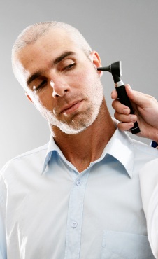
Use of Otoscope for Diagnosis of an Earache:

Otitis Externa: An otoscope can be used to diagnosis otitis externa as the ear canal will appear inflamed and swollen. Thick discharge may be present and the insertion of the scope into the canal will cause pain to the patient.

Otitis Media: An otoscope can be used to diagnosis otitis media as the physician examines the ear drum. Fluid bubbles and air may be present inside the ear drum and it may look red and inflamed.

Bullous Myringitis: An otoscope can be used to diagnosis bullous myringitis by inserting the scope to examine the ear drum. Small blisters filled with fluid may be present.

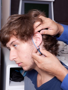
Hearing Tests for Diagnosis of an Earache

Hearing tests may be recommended for patients who have had reoccurring earaches or ear infections. They may also be used for children with a speech delay as this can be connected to an earache. During a typical hearing test, the patient may wear a pair of headphones and listen to various tone depths. If they are unable to hear some or all of the tones, they may have an ear infection.

Laboratory Tests for Diagnosis of an Earache

Drainage samples from the infected area are sometimes sent to the hospital laboratory to check for signs of bacteria. Laboratory tests are not typical for ear infection patients and are reserved for those individuals who do not respond to other treatments.

Treatment Options

The treatment of earaches commonly requires the use of antibiotics of self care remedies. Occasionally, a patient may need a surgical procedure performed to completely cure the damage done by an ear infection or ear trauma.

Home Self-Care for Earaches

Although ear infections should be diagnosed and treated by a physician, the uncomfortable symptoms caused by the infection can be treated at home. The use of over the counter pain relievers such as ibuprofen or acetaminophen can be used to reduce pain accompanied by the earache. A warm compress can also be placed against the infected area to help with pain control.

Medical Treatment for Otitis Externa

The majority of cases of otitis externa are cured with the use of prescribed eardrops for a period of seven to ten days. These eardrops contain a steroid and antibiotic to fight off infection and reduce inflammation and swelling in the ear. The drops are placed in the ear while the individual in lying on their side. The patient should remain on their side for a minimum of five minutes to allow the drops to enter the ear canal. An oral antibiotic may also be prescribed to keep the infection under control through treatment. During the treatment, the ear canal should be kept completely dry. Ear plugs should be worn for bathing, showering and swimming during this time.

Medical Treatment for Otitis Media

There are several treatments available for otitis media. Oral antibiotics are typically given to adults who acquire the condition for quick treatment. Observation is usually the only treatment needed for infants and children as most cases of otitis media in children will treat itself over a period of one to two weeks. For more severe cases of otitis media in children, follow up care may be needed resulting in antibiotics to cure the ear infection. Over the counter pain medications may be recommended to reduce the pain accompanied by the earache. A humidifier may also be used to moisten the air.

Medical Treatment for Bullous Myringitis

Treatment for bullous myringtis may include ear drops, oral antibiotics and symptom related pain medications. Bullous myringtis earaches usually resolve themselves in one to two days of treatment. It’s important not to stop the treatment before all the antibiotics are taken as the infection may still be present even if the symptoms are gone. A follow up may be recommended as well as a hearing test if symptoms do not subside. Your physician may refer more severe cases to an ear, nose and throat specialist for future examination.

Conditions

Amnesia

One of the most terrifying injuries that can happen to a person involves suffering from amnesia. Imagine losing your entire memory, all of your life’s happiness and all of the pain; or imagine not knowing who your family is, or even who you are. Amnesia can affect anyone, at any age and at any point in his or her lives. Amnesia can be either the loss of short term memories or long term memories; or in some cases, even both. In addition, patients may have difficulties imagining what their futures may hold, as they may not be able to recall the plans or dreams they had before the onset of the amnesia. In general, there are two main forms of amnesia: Anterograde Amnesia and Retrograde Amnesia. A patient can suffer from either form, or in the worst cases, both forms.

Anterograde Amnesia

With Anterograde Amnesia, the patient loses the ability to create and retain new memories after the incident that caused the onset of the amnesia. This can also lead to difficulties recalling events that have happened in the most recent past, while memories of their distant past and long term events remain in place. Anterograde Amnesia remains somewhat of a mystery to physicians and neurologists, as the process of retaining exact memories is not yet widely known.
Memory Loss
It is believed that there are two main causes of Anterograde Amnesia: drug or alcohol induced or as the result of a traumatic brain injury; however the condition has also been known to be the result of an emotional disorder or a shocking event. In one of the most common forms of Anterograde Amnesia, alcohol induced blackouts, studies have shown that with rapid increases in the blood alcohol concentration within a short period of time can severely impair the brain’s ability to transfer the short term memories into long term memories for storage and the ability to later retrieve them; this is what happens when you hear someone discussing how they are unable to recall the events of the night before.

Drugs and Alcohol Consumption

By drinking large amounts of alcohol either in a short period of time or on an empty stomach, one can have difficulty remembering the events of the time frame in which they were intoxicated, however once they have reached sobriety, their ability to create and retain long term memories is restored in full.

In addition to rapid alcohol consumption, using drugs that are considered benzodiazepines including midazolam, flunitrazepam, triazolam, nimetazepam and temazepam are known to have profound effects on one’s memory functions.

Similar to the effects of rapid increases in blood alcohol concentration, taking an excess of these drugs can impair the brain’s ability to create short term memories and transfer them into long term memories. In cases where the amnesia is either drug or alcohol induced, the patient can easily recover from it, usually after waking from their induced state.

Retrograde Amnesia

Individuals suffering from Retrograde Amnesia are often found to be unable to recall events that took place before the amnesia set in. In the majority of cases, Retrograde Amnesia is caused by a traumatic brain injury, possibly resulting from a fall or a car accident.

Those who suffer from Retrograde Amnesia can recover their lost memories, although some of the memories that one would had closest to the onset of the amnesia may never be fully restored. It has been described as feeling like the hours leading up to the onset of the amnesia feel more like a dream than actual reality. Currently there is no cure for Retrograde Amnesia, although doing activities designed to “jog” one’s memory may help them remember some skills or events, but not their entire life before the onset.

Types of Memory

There are two types of memory that are most likely to be affected by amnesia: procedural memory, which is the ability to remember how to do certain skills such as tying your shoes or driving a car; the other, declarative memory, is the part of the memory that retains information regarding events that have happened in one’s life. It is also the part of the memory that retains information learned in school, such as history.

Types of Amnesia

Post-traumatic amnesia is commonly described as a confused state as the immediate result of a traumatic brain injury. During an instance of post-traumatic amnesia, the individual is unable to recall events that happened after their injury took place. They may not know their name, where they are or when in time it is. While they are suffering from the effects, new memories cannot be stored. Once they begin to regain their memory, the post-traumatic amnesia is considered to have resolved itself.

Psychogenic amnesia, which is more commonly referred to as dissociative or functional amnesia, is diagnosed when one has the presence of retrograde amnesia but does not have the presence of anterograde amnesia. Individuals suffering from psychogenic amnesia cannot remember things about themselves like their own identity, but they are able to retain new information and perform daily functions as they normally would. At times, individuals can lose the ability to read or write, and will have to re-learn these skills.

Repressed Memory & Childhood Amnesia

Repressed memory is a form of amnesia where an individual is unable to recall the events that are traumatic or stressful in an individual’s life. For example, a rape victim may subconsciously block out the circumstances surrounding their attack.

While the memory is stored in a person’s long term memory, the psychological defense mechanisms in their brain will not allow them to remember it. The patient will still have the ability to learn new information, and as time passes, they may begin to recover all or some parts of the memory they had previously blocked out.

Childhood amnesia, also referred to as infantile amnesia, is best described as the inability to remember events from a person’s own childhood. This can refer to traumatic events or even just remembering activities or vacations taken with one’s family. The majority of patients have reported having difficulties remember events that occurred from birth to the age of four years.

Alternative Names

Both Anterograde Amnesia and Retrograde Amnesia are most commonly referred to as either basic amnesia or memory loss.

Additional Causes

In addition to head trauma and drug interaction, amnesia can also be a result of surgeries performed on the brain. At times neurosurgeons may have to remove a part of the brain in order to prevent or treat a more serious disease, or the parts of the brain can become damaged during surgeries. An individual suffering from a brain tumor that needs to be removed runs the risk of having one of the parts of the brain that is a part of the memory process either damaged or removed, causing either Anterograde or Retrograde Amnesia.

Symptoms

The easiest symptom of amnesia to recognize is memory loss; whether it is only a part of the memory or total loss, or whether it is a temporary setback or a more permanent loss. Individuals may also show difficulty in retaining new memories and information. One may also experience false recollections of events, at times inventing memories or confusing multiple events with one another. In addition, an individual who is suffering form amnesia can experience neurological problems including tremors and seizures, as well as appear confused or disoriented.
Memory Loss

Risk Factors

If one has experienced certain injuries or events, they may be at a higher risk for amnesia. These include, but are not limited to, brain surgery and trauma or injury to the head; abusing alcohol and strokes or seizures. And while an individual can live with amnesia, the complications from dealing with it can impact ones daily life. Being unable to recall specific memories can lead to problems in social settings, at work or school and at home.

Some may never recover their lost memories. And those with the most severe cases of amnesia may require constant supervision or be required to live in an extended care home where they can be monitored, as even completing the smallest tasks can become impossible to do.

Prevention Tips

Unfortunately, it can be impossible at times to completely prevent amnesia. No one can predict when they may experience a traumatic injury such as a car accident; or when they may fall victim to a vicious attack such as a rape. However, the following tips are recommended to decrease your chances of suffering from amnesia in the event of an accident: wearing a helmet if participating in dangerous sports or bicycling; wearing your seat belt when in an automobile; avoiding the excessive use of drugs or alcohol.

For those who have a head injury, make sure to get treatment as soon as possible after the incident. And if one feels that they are suffering from a brain aneurysm or a stroke, seek medical attention immediately.

Treatment

The treatment of amnesia will vary depending on the type and cause of amnesia, as well as how each individual person’s body reacts to the cause. One treatment for those who are suffering from amnesia as the result of a traumatic event is cognitive rehabilitation.

The individual would need to be surrounded by love and support, as well as counseling to overcome what happened to them, although recalling such memories can do more harm than good. Psychotherapy and hypnosis is another option, as it can aid in recovering the lost memories. In the case of drug or alcohol abuse, emotional support as well as counseling to determine the root cause of their addiction is needed.

If the amnesia is caused by a condition that affects the brain such as an injury or Alzheimer’s, the treatment will be much more medically based. In the case of physical damage to the brain caused by injury, it may very well be impossible to treat with anything other than time and/or therapy.

Some patients find that they recovered their memory once the brain has had time to adapt to the trauma, while others have unfortunately found the loss to be permanent.

Treating Alzheimers & Living with Amnesia

In the case of a disease such as Alzheimer’s, the best course of treatment for amnesia is to attempt to prevent any additional memory loss. What is gone will most likely never return as an individual advances through the stages of the disease.

There are prescription options that an individual can take that work to either protect the brain itself or slow down the rate at which the memory is lost. However, as with most prescriptions, side effects can be significant and may include nausea and vomiting, loss of ability to sleep and loss of appetite. In most instances, the drugs merely slow down the inevitable.

While living with amnesia is not the best scenario for anyone, as medical technology advances doctors may be able to determine a more specific root cause of the disease, and in turn come up with a cure. But in the meantime, being there and supporting those who are dealing with amnesia and helping to restore their precious memories is a key part of helping those who are living with the disease do just that: Live.

Conditions

Allergies

Allergies, estimated to afflict one out of every five Americans, are abnormal reactions to normally harmless substances in the environment called allergens. Upon exposure to an allergen, the body’s immune system mistakenly identifies it as a threat and takes defensive action against the presumed invader. The immune system’s inappropriate attempt to protect the body gives rise to allergy symptoms, which can affect multiple organ systems and which range from the mild to the life threatening.

Symptoms

Allergy symptoms vary according to the nature of the allergen and the way in which it encounters the body, and can be roughly divided into three categories that are defined by the manner in which an allergen enters the body.
Allergy Symptoms

Airborne Allergens

Airborne allergens like dust, pollen and pet hair travel through the air and come into direct contact with the eyes, nose, throat, sinuses and lungs. This contact gives rise to a constellation of symptoms including sneezing, nasal irritation, itching and watery eyes and runny nose. Allergic rhinitis, commonly known as hay fever, falls into this category, with symptoms that occur seasonally or year round in response to various pollens and spores emitted by trees, grasses and weeds. Pet allergies can be triggered both by airborne exposure and by contact, and the airborne component causes many of the same symptoms as hay fever. Animal fur is not the culprit. Instead, symptoms are caused by exposure to minute flakes of skin, or dander, shed by the animal.

Many of the symptoms associated with airborne allergens, while uncomfortable, are relatively mild, but serious symptoms do occur. Severe symptoms can begin with wheezing and shortness of breath and escalate to full-blown asthma attacks and difficulty breathing caused by swelling of the throat.

Ingested Allergens

Food, medication and insect stings are all capable of producing allergic reactions.

Food allergies can cause stomach pain, vomiting, diarrhea and itching or swelling of the skin. As digestion progresses, symptoms may spread from one organ system to another, beginning with itching in the mouth as the food is taken in, followed by pain, vomiting or other abdominal symptoms as the food moves through the system. As digestion continues, food is broken down and its components are released into the bloodstream. In this way, allergens may be released into the blood and transported throughout the body.

Since the dissemination of the allergen is so widespread, symptoms can affect the skin in the form of hives or eczema, the digestive system itself or the circulatory system as a whole. In the last case, the result can be anaphylaxis, a sudden drop in blood pressure than can be fatal if not treated immediately.

Medications and insect stings give rise to allergy symptoms that can simultaneously affect several organ systems. Symptoms can appear immediately upon exposure or their onset can be delayed. They may include itching or hives, difficulty breathing, swelling of the tongue or throat, stomach pain, vomiting and cramps, all symptoms of anaphylaxis. Loss of consciousness and coma may occur as the reaction continues. Symptoms may appear to be diminishing, only to return in force even if there has been no subsequent exposure to the allergen.

Contact Allergens

Allergic reactions can occur when the skin is exposed to certain substances, including poisonous plants, latex and animal saliva.

Symptoms resulting from contact exposure most often begin with itching and redness of the skin at the point of contact, followed by the development of a rash, blisters or hives. Symptoms of exposure to poisonous plants generally subside after a week or two, but may persist in particularly sensitive individuals for up to 30 days. While uncomfortable, symptoms do not generally become more serious, although infection can occur at the affected site. However, serious respiratory symptoms can result from the inhalation of smoke from burning poisonous plants like poison ivy or poison oak. This mode of exposure usually necessitates extensive medical treatment.

Latex allergy chiefly occurs among health care workers, where symptoms of exposure are typically limited to a localized rash or irritation called contact dermatitis. More serious symptoms, including anaphylactic reactions, can occur following abdominal surgery and medical and dental procedures that expose patients’ mucous membranes to latex gloves.

Causes

Once the immune system marshals its defenses, all allergies follow the same physiological pattern regardless of the allergen involved.

  • First,  person is exposed to an allergen through the skin, the respiratory system or the digestive system, and the immune system identifies the allergen as a threat.
  • This initial exposure does not cause any allergic symptoms, but sets the immune system to work against the invader.
  • Certain white blood cells begin to produce an antibody called Immunoglobulin E, or IgE, whose job it is to bind the allergen.
  • IgE circulates throughout the body and attaches itself to mast cells, which play a key role in the inflammatory process and are rich in histamine.
  • At this point, the immune system has been sensitized to the particular allergen.
  • The next time the immune system encounters the allergen, it binds the allergen and the mast cells release histamine and a broad arsenal of other chemicals that produce inflammation.
  • This time around, the release of those inflammatory agents, known collectively as “mediators,” results in symptoms.

Whether symptoms are localized or general depends on the specific allergen and the person involved, and whether an allergic reaction is mild or severe depends to some extent on the extent and suddenness of the release of mediators. The presence of great numbers of mast cells in the respiratory and digestive tracts explains the frequent occurrence of allergy symptoms in those locations.

Risk Factors

While the physiological causes of allergy are well understood and relatively straightforward, risk factors involve a complex interaction of genetics, environment and individual history and susceptibility.

Heredity

Heredity plays a strong role in the probability that a person will be subject to allergies. Children in families where both parents are allergic have at least a 60% chance of having allergies. Such children are more likely to be sensitive to more allergens and to have allergies that are more severe than children who do not have an allergic parent. In general, this genetic predisposition does not translate into the inheritance of sensitivity to a specific allergen, but studies of identical twins have found a 70% likelihood that the twins will suffer from the same allergies.
Allergy Risk Factors
In children, the incidence of allergy is higher in boys, although this imbalance tends to disappear in adults.

Racial and ethnic factors have also received some attention from researchers, particularly in light of the higher distribution of asthma in the African American population, but results have been inconclusive, especially because of the difficulty of separating environmental and genetic influences.

Environment

Almost by definition, the environment is a risk factor for allergy. After all, allergies do not exist without allergens and allergens come from the environment, whether in the form of pollen, food, medicine, pet dander or poison ivy. Some environmental risk factors are not as self-evident.

Public health researchers have noted a number of epidemiological trends that suggest a broad environmental basis for the development of allergies.

  • In the Western world, the incidence of allergies has approximately tripled between 1980 and 2010.
  • Allergies are much more common in the industrialized world than in the developing world.
  • Immigrants from the developing world to the industrialized world show a greater risk of allergy, and that risk grows along with their time in the industrialized world.
  • Children in larger families are less likely to develop allergies than those in smaller families.

The generally accepted explanation for these trends, the “hygiene hypothesis,” holds that the immune system benefits from exposure to a wide variety of irritants and bacteria and thus becomes more tolerant of foreign substances. In the absence of that kind of stimulation, the immune system is more likely to react to a normally harmless substance like pollen.

Other environmental factors contribute to the risk of developing allergies, chiefly by serving as irritants. The likelihood of allergic reactions, especially in children, increases with exposure to air pollution and tobacco smoke.

Personal Factors

Some risk factors are associated with a person’s individual history or with other medical conditions.

A person who has been exposed to allergens in childhood is more like to develop allergies in later life, and more likely to react to the allergen to which the child was exposed. Continued exposure to known allergens increases the risk that a person will become allergic to them.

Frequent ear, nose and throat infections in childhood also correlate with increased allergy risk, as does treatment in early childhood with broad-spectrum antibiotics and the use of antibacterial cleaning products in the home. There is also evidence that infants who are breast-fed and are not given cow’s milk or solid foods for the first six months of life are less prone to allergies later.

Among medical conditions, the presence of nasal polyps, asthma and skin sensitivity are all conditions that correlate with the development of allergies. People with high levels of IgE in the bloodstream are also more likely to develop allergies.

Prevention

Allergy prevention begins with avoiding exposure to the substances that trigger allergic reactions. Therefore, preventive strategies vary according to the allergens and irritants involved.

Pollen and Mold

  • Close windows at home and in the car.
  • Avoid outdoor activity in the early morning and late afternoon, when pollen counts are high.
  • Use air conditioning to filter pollens from the air, and change filters regularly.
  • Keep indoor spaces ventilated in order to cut down on moisture.
  • Hard surfaces are easier to clean and less conducive to mold growth, so avoid rugs and carpets, especially in the kitchen and bath.
  • Keep indoor plants to a minimum, as they can provide homes for mold.
  • Avoid drying clothes outdoors where they tend to accumulate pollen.

Pet Dander

  • Limit or eliminate the pet’s access to bedrooms.
  • Wash the pet frequently, in order to remove dander and pollens that can collect on the pet’s fur.
  • Use air filtration machines, either central systems or room units, preferably with HEPA filters.
  • Foods
  • Read food labels and question restaurant staff about ingredients.
  • Learn to recognize early symptoms, since allergic reactions to food can happen quickly and may require immediate intervention.
  • Introduce solid foods to infants gradually, beginning when the child is at least six months old.

Poisonous Plants

  • Learn to identify plants that produce allergic reactions.
  • Wear gloves and long pants and sleeves when encountering potentially poisonous plants.
  • Avoid burning plant materials, especially if any are suspect.
  • Medications
  • Be sure to inform health care providers of any known allergies and of any family history of medication allergies.
  • Wear a medical alert bracelet that identifies the allergy if you cannot communicate.

Diagnosis

The two medically accepted avenues of diagnosis are skin testing and blood testing, although a vast array of unproven and generally invalid methods competes with the established diagnostic tools.

Skin Testing

In skin testing, the back or arm is pricked with a metal or plastic device that makes a series of tiny punctures in the skin. Small amounts of the proteins associated with suspected allergens are applied to the skin and mapped according to their placement. If an allergy to the particular substance is present, the individual site will redden or develop a hive. Skin testing has demonstrated a high degree of diagnostic accuracy in all areas except for food allergies, where blood testing provides results more clearly related to specific allergens.

Blood Testing

Blood testing is more complicated and expensive than skin testing, but is sometimes preferred for patients who have had severe anaphylaxis as well as for patients who have recently taken antihistamines or who have diseases of the skin. It does allow for greater accuracy in pinpointing food allergies.

In blood testing, a small amount of blood is drawn and analyzed for the presence of IgE antibodies that are specific to a particular allergen. High levels of allergen-specific IgE antibodies indicate the likelihood of an allergic reaction to the allergen.

Unproven Tests

Unproven tests greatly outnumber the two tests that have proven medical validity. These procedures include measurement of changes in the electrical resistance of the skin, changes in muscle strength on exposure to a suspected allergen and changes in pulse rate after eating a suspected allergenic food. Other unconfirmed methods are the microscopic examination of blood cells in contact with an allergen and the analysis of samples of hair and tissues for trace amounts of chemicals alleged to be toxic. No evidence supports the ability of these tests to provide any benefit to the patient, diagnostic or otherwise.

Treatment

Allergy Treatments
Prevention is the first line of allergy defense, but it is impossible to prevent all exposure to allergens, especially to allergens like pollen that are ubiquitous in the environment. There are many available treatment options, from home remedies to sophisticated immunotherapy, and modern treatments have proven to be extremely effective.

Over-the-counter Medications

Antihistamines – As their name suggests, antihistamines work by blocking histamine receptors in the body. They have a long history of use in treating allergy symptoms and can be taken when needed for relief of sneezing, runny nose and watery eyes. Early antihistamines were known to cause drowsiness, but there are now drugs available, including loratadine, that provide relief without the troublesome side effect, Antihistamines are available as pills, liquids and nasal sprays and as eye drops can help treat itchy eyes.

Decongestants – Decongestants shrink blood vessels and swollen tissues in the nose, helping to decrease their tendency to produce fluid. Although they can be taken as needed in response to symptoms, long term use of decongestants as nasal sprays can lead to a worsening of symptoms once the decongestant is stopped. They tend to raise blood pressure and may be contraindicated in people with hypertension. Decongestants are available as pills, nasal sprays and eye drops, and by prescription in combination with other allergy medications.

Combined medicines – Medicines are also available that combine two different active ingredients. Most commonly, these consist of an antihistamine and a decongestant, although other preparations combine allergy medicines with pain medications, asthma medications or other ingredients.

Prescription Medications

Bronchodilators – Bronchodilators work by opening the airways in the lungs and allowing mucus to move more freely. They are used to treat the symptoms of asthma and come in short-acting forms for immediate relief and long-acting forms that last for up to 12 hours. Bronchodilators can raise blood pressure and increase the pulse rate. They are available as inhalers, as they must be inhaled into the lungs in order to be effective.

Leukotriene modifiers – Leukotrienes are produced by the immune system and contribute to inflammation in allergies and asthma. Leukotriene modifiers suppress their production. Side effects are uncommon, mainly involving stomach upset or pain, but the medication must be taken daily in order to have the desired effect. Leukotriene modifiers are only taken orally.

Mast cell stabilizers – Mast cells are the histamine-rich cells to which IgE attaches in the allergic process, and stabilizer drugs prevent the cells from releasing histamine. They may also have some anti-inflammatory effects and are useful in preventing asthma symptoms. In allergy treatment, they are used as a preventive medication taken prior to exposure to a known allergen. Mast cell stabilizers may take up to a month to reach full therapeutic potential. They can be taken orally or used as inhalers or nasal sprays.

Steroids – Steroids chiefly act by suppressing inflammation and are among the most versatile of allergy medicines. They relieve asthma symptoms, as well as nasal congestion, runny nose and sneezing. Steroidal creams can be applied to the skin to treat symptoms caused by contact with allergens. Side effects of steroids are as varied as their uses and include high blood pressure and weight gain in the short term and cataracts, diabetes and muscle weakness in long-term use. Steroids are available as pills, nasal sprays, eye drops and topical creams. When taken orally, steroids are generally prescribed as a course of treatment lasting at least several days, but they are also effective when used as needed.

Epinephrine – Epinephrine, also known as adrenaline, is used as an emergency treatment for anaphylaxis. It is injected immediately upon the appearance of anaphylaxis symptoms, including itching, swelling of the mouth or tongue, trouble breathing and nausea. Side effects can include hypertension, heart palpitations, racing pulse, anxiety and headache, but none of these side effects contraindicates the use of epinephrine in a potentially deadly onset of anaphylaxis. Epinephrine is only available in injectable form and comes in a small syringe containing a premeasured dose. The EpiPen is perhaps the best-known brand. A variation that includes a second dose, the Twinject, is designed to manage the relatively common situation in which symptoms of anaphylaxis have subsided only to reappear several minutes later.

Immunotherapy

Immunotherapy does not cure allergies, but it is the closest medicine has yet come to providing permanent relief.

Immunotherapy consists of a series of injections given in the upper arm for a period of several years. Initial treatment is given once or twice per week and, if successful after the first several months, once or twice per month for the duration of treatment. The shots consist of gradually increasing doses of a specific allergen in the hope that the patient will become desensitized and develop something akin to immunity to the allergen used.

Immunotherapy has proven to be a safe and effective treatment for allergic asthma and for allergies to pollen, pet dander, molds and insect bites. It is not considered an effective treatment for food allergies. Side effects are rare, although there is a small risk of a severe reaction to the allergen injected.

Other delivery methods have been used with some success outside the United States, including oral and intranasal administration of the dose of allergen. In addition, attempts have been made to shorten the time necessary to build up to a maintenance dose by reducing the interval between allergen exposures to as little as several hours. This strategy is used only in a hospital setting as it entails greater risk of a severe reaction to the allergen.

Home and Herbal Remedies

Home treatments are capable of relieving a number of allergy symptoms. For nasal congestion and symptoms of hay fever, nasal irrigation washes irritants from the nose and sinuses. A bulb syringe or neti pot can be used to introduce a saline solution to the nose.

For insect stings, aloe vera or a paste of water and baking soda can relieve pain and itching at the site of the sting.

Many herbal remedies have been proposed as allergy treatments, including goldenseal, stinging nettle and butterbur. Although a comprehensive 2006 Mayo Clinic study of herbal and other nontraditional approaches found little evidence of their efficacy, two recent European studies of butterbur concluded that it is as effective as an oral antihistamine in alleviating hay fever symptoms, and that it does so without causing drowsiness.

Unproven Remedies

Alternative therapies like homeopathy, kinesiology, naturopathy and probiotics find little support in the medical literature and have not been shown to be effective when studied individually.

Acupuncture stands out as the one exception to those disappointing results, although its promise derives from two small studies. The first showed an improvement in hay fever symptoms in all 26 of the patients studied, and did not report any side effects or adverse reactions. The second study involved 76 patients and reported that two sessions of acupuncture eliminated symptoms in over half of those participating.

Resources

CDC.Gov
EverydayHealth.Com
MayoClinic.Com

Conditions

Thinning Hair

Thinning Hair and Hair Loss

Hair loss in any form is a cause of distress for most people. Many assume that thinning hair, or loss of hair is simply an unavoidable aspect of aging. Others assume it is determined by heredity and therefore, cannot be avoided. It is important to note that there are numerous causes of thinning hair and a variety of treatments available to address this issue. Often thought of as a problem primarily plaguing men, the issue of thinning hair and/or hair loss affects numerous people, both male and female, sometimes at a fairly young age.
Thinning Hair

Hair Loss Terms

  • Alopecia is the common medical term used to describe the loss of hair either on the head, or on any part of the body.
  • Male Pattern Baldness refers to the loss of hair from the front and central part of the head, with hair usually remaining on the sides and back of the head.
  • Female Pattern Baldness is a term used to describe the thinning of hair on a woman’s head. Typically this is an overall thinning, not specifically affecting a woman’s hairline, with the primary spot for thinning being on the top of the head.
  • Effluvium is a term that is used in combination with other medical terms to describe the type of hair loss. The word by itself means a shedding of hair.
  • Trichotillomania is a condition where a person, either child or adult, is in the habit of pulling out normal, healthy hair.

How Hair Grows

There are three basic phases of hair growth in normal hair follicles. The growing phase is called anagen. This is the longest phase and can last from two to eight years. Next is a transition phase that represents the time when the hair has stopped growing and is entering the resting phase. This period lasts for about two to four weeks, as the hair follicle shrinks, and the hair stops growing, but remains attached.

This transitional phase is called catagen. The last phase is the resting phase, or telogen. This phase lasts from two to four months. After the resting phase is over, the hair naturally falls out, as new hair grows in to replace it. On any given day, it is estimated that a person with a normal, healthy head of hair has up to 100 hairs that have finished the telogen phase and fall out.

Symptoms of Hair Loss

The symptoms for hair loss are quite obvious in most instances, particularly for men. A visibly receding hairline is often the first indicator. For women, it may take some time for the thinning to be noticeable. In fact for some women, the first indicator is a noticeable increase in the quantity of hair coming out in a brush, or collecting in a drain after the hair has been washed. In some instances, a woman may be alerted to the condition by her hair dresser. In any case, whether male or female, the loss will eventually be visible to the eye.

Causes of Hair Loss

Hair loss can be caused by a vast number of things(opens PDF). In many instances, a person’s head of hair can reflect a person’s overall health.

In some instances, the loss can be caused by a known drug, the most obvious being chemotherapy. It is important to note that other drugs can also cause hair loss, including some drugs used to treat common ailments like high blood pressure or depression. This type of hair loss is sometimes referred to as Anagen Effluvium, as in many cases, the hair falls out or is disrupted during the anagen or growing phase. Anagen Effluvium is also found from exposure to chemicals like mercury.

Poor nutrition can be blamed for some cases of hair loss. From B5 (Pantothenic Acid) to B2 (Riboflavin), to a deficiency in essential fatty acids, people who are not adhering to a well balanced diet may experience some form of hair loss. Essential vitamins and minerals are vital to healthy hair.

Physical and emotional stressors can also cause a person to experience thinning hair. A severe illness or a sudden loss of weight, are both common causes of hair loss. Some cases of hair loss can be directly attributed to thyroid issues or anemia.

In some instances, hair loss is permanent, and may be caused by inflammation brought on by a variety of skin conditions. This type of hair loss is called Scarring Alopecia, and typically occurs when, as the name implies, hair follicles are scarred.

The most common cause of baldness or thinning hair is frequently attributed to three things: hormones, heredity and aging. Although this type of balding can begin as early as the teen years for men, it is more frequently observed with age, and seems more likely to have a strong connection to heredity.

Risk Factors

When reading through the causes listed previously, there are some obvious indications of those who are at higher risk for hair loss and thinning hair. Some factors, like heredity, are beyond a person’s control. Others, like diet and stress can be addressed and approved either alone, or with help.

Preventing Thinning Hair

Thinning Hair Prevention
First and foremost, pay attention to your overall health. Consume a well balanced diet and take a multi vitamin. If you regularly experience stress, try to incorporate a daily de-stressor into your schedule. Consider activities like yoga, meditation, or virtually any kind of physical exercise. These are all proven stress reducers, and can play big part in a person’s overall well-being, which in turn plays a part in your hair health.

For women or men with long hair, refrain from putting your hair up in braids or tight pony tails, as the stress of sustained pulling on the hair can cause hair loss. Be cautious of using harsh chemicals in any aspect of your life, but particularly in hair dyes or other hair treatments. Consider incorporating any of the herbal remedies shown to promote healthy hair. (See Treatment Options).

Testing and Diagnosing

Because there are a wide variety of conditions that can contribute to hair loss, tests performed during a diagnosis are also widely varied. From a nutritional analysis, to blood tests, to the microscopic study of a sample hair shaft, a patient may undergo any number of types of tests to rule out, or diagnosis underlying health conditions. In some rare instances, for some conditions, a skin biopsy might even be performed.

Treatment Options

Depending on the underlying cause of hair loss, and if it can definitively be diagnosed, treatment options are varied.

For patients experiencing hair loss that is attributed to aging or heredity, there are a wide number of prescription drugs currently being prescribed. Minoxidil, also known as Rogaine, is widely used. It comes as a topical solution, and is applied directly to the scalp. Finasteride, also known as Propecia, is an oral medication prescribed for men only. It works to block the negative effects of male hormones on hair loss.

For Minoxidil products, the most common side effect is an itchy scalp, often treated with a simple dandruff shampoo. For Finasteride, side effects may include erectile dysfunction, or decreased libido, but these are seen in less than 20% of users.

Surgical hair transplantation is another viable option. In this procedure, healthy hair follicles are moved from one area of the scalp to an area that is balding. The side effects associated with this type of treatment are pain, swelling and some bleeding, all diminishing with time. A patient may also experience headaches, which normally can be treated with over the counter pain relievers.

For some patients, simple natural treatments are available and recommended, especially to treat hair loss that is attributed to diet or stress. Vitamins and exercise are two obvious, but very key treatment options.

Herbal Options

There are a number of herbal remedies recommended for the treatment of thinning hair or baldness. These remedies generally fall into two categories: those taken internally and those applied topically. Topical remedies might include a mixture of essential oils like thyme, rosemary, lavender and cedarwood, applied directly to the scalp. Another commonly used topical treatment is coconut oil.

This treatment is particularly popular with women as it acts as a natural conditioner. Aloe Vera has also shown promise in promoting thick, healthy hair. Burdock Root Oil is another popular treatment, often added directly to your existing shampoo, making it a particularly easy one to try.

Herbal remedies taken internally include herbs used to improve blood circulation, including gingko biloba. Horsetail is another herb that is used to promote circulation and blood flow. It is thought to increase hair strength, and is frequently taken as tea. Other herbs, like green tea, are believed to block enzymes linked to hair loss. Green tea may be found either as a drink, or in capsule form. When trying any herbal treatment, it is important to watch for any allergic reaction, as these types of reactions are the most likely side effects.

Trichotillomania

For patients diagnosed with Trichotillomania, treatment may include Cognitive Behavioral Therapy (CBT), performed by a trained psychologist. Generally speaking, this involves an attempt to determine the exact triggers causing a patient to pull out hair, and then teaching the patient ways to respond differently to those triggers. Some patients respond best to a combination of CBT and medication, such as one of the standard medicines used to treat depression. Others have been helped with dietary changes, hypnosis, yoga, or other natural remedies.

Scarring Alopecia

The most difficult type of hair loss to treat is the Scarring Alopecia variety. Because it is difficult to treat, most physicians will attempt aggressive treatment that may include corticosteroids in topical cream form or in injection form, or any of a variety of prescription drugs. Once a patient with this form reaches a time where the hair loss seems to have ceased, if any healthy hair follicles remain, the patient may be a good candidate for transplant surgery.

As with the treatment of any physical condition, it may take some time to determine what treatment protocol works best for each individual person. What works for one person may not produce the same results in the next. Research is constantly under way, investigating new possibilities for the treatment of hair loss.

Next Steps for Treating Hair Loss

For any person dealing with thinning hair or hair loss, it is important to realize that there are number of possible causes, and a number of available treatments. Taking time to examine your overall health and diet is the best first step.

For those who feel that heredity is leading them down a path of male or female pattern baldness, taking a pre-emptive strike may be the best possible path. Of course this includes diet and stress management, but it may also represent a great opportunity for the introduction of any of the numerous natural remedies associated with promoting hair growth.

Conditions

Leucorrhoea

Leucorrhoea, or, as it is also known as, leukorrhea or leucorrhea, is a normal and common condition in which discharge from the vagina becomes changes in color or consistency. This discharge is often caused by inflammation or infection within the vagina or cervix. Irritation of the membrane lining can also cause leucorrhoea to occur. This discharge may become thick or foul smelling, or increase in quantity around puberty or during sexual arousal. It is referred to as either cervical leucorrhoea or vaginal leucorrhoea, depending where the discharge originates.

Symptoms

Leucorrhoea itself is a natural increase in vaginal discharge but it can become bothersome and disruptive to the normal life of a female. Many factors can contribute to the cause, and some symptoms may be indications of a more serious condition. Care should be taken to report any unusual changes to your physician when applicable.

leucorrhoea symptoms This discharge may be like any of the following:

  • Dark, bloody, yellow or white in color
  • Foul or sour smelling
  • Thick, watery, sticky or frothy

Other symptoms can include:

  • Pain during or after intercourse
  • Spots on undergarments from discharge
  • Lower back or abdominal pain
  • Leg pain, specifically calf and thigh
  • Frequent urination, especially burning during urination or passing little urine
  • Fatigue or weakness
  • Irritability and lack of concentration
  • Upset stomach such as diarrhea or constipation or increased gas production
  • Frequent headaches
  • Black or dark circles under the eyes
  • Itching in the vaginal and surrounding areas, area may become inflamed or reddened

Causes

Many causes of leucorrhoea exist, the most frequent one being hormonal imbalance, but may be caused by infection or irritation of the membranes lining the genitals.

Other causes can include:

  • Spermicidal creams, ointments and jellies that are used as contraception which can irritate the lining or strip the mucus
  • Certain medical conditions such as diabetes and anemia
  • Bacterial or fungal infections
  • Irritation caused by IUDs (intra uterine device used for contraception)
  • Injury to the cervix or vagina caused by sexual activity, muscle injury, childbirth or miscarriages
  • Unhygienic conditions that lead to the accumulation of bacteria within and around the vagina
  • Sexual intercourse
  • Sexually transmitted diseases. Gonorrhea often causes a marked discharge.
  • Menstrual changes such as onset of puberty, menopause or changes associated with pregnancy

There are also several classifications of the discharge that have specific names and symptoms.

Cervicitis

Cervicitis is often indicated when lower back pain is present. Cervicitis is caused when the uterine cervix becomes inflamed. This may occur due to infectious causes such as sexually transmitted diseases but non infectious causes as possible. The most common non infectious causes are allergic reactions to spermicidal products or lubricants, latex condoms, contraceptive diaphragms, and intra-uterine devices (IUDs).

Monilial Vaginitis

Monilial Vaginitis, also known as a vaginal yeast infection or candidiasis, occurs most often in pregnant or diabetic women, or also in women who take birth control pills or some antibiotics. Hormonal changes and oral contraceptives have been shown to change the pH in a woman’s body, which can lead to yeast infections. Other factors may contribute to vaginal yeast infections, often by wearing dirty or damp undergarments for long periods of time and unsanitary conditions.

Trichomonal Vaginitis

Trichomonal Vaginitis, also called trichomoniasis or trich, manifests as a yellow and sometimes frothy discharge along with itching. It is commonly caused by sexually transmitted diseases, but can also be caused by poorly cleaned sexual toys, wet towels and unclean or damp undergarments worn for long periods of time. Symptoms can appear within 3 to 28 days after infection and may be anywhere from a mild discomfort to severe complications.

Pregnant Women

Women who have become pregnant may face an increased risk of premature labor if they are infected with trichomonal vaginitis. The risk of being infected by other sexually transmitted diseases such as Chlamydia also is increased due to this type of leucorrhoa. Sexual partners may need to be treated for this as well to prevent recurring infections.

Risk Factors

The primary risk for leucorrhoea is it becoming a chronic condition or severe infection. This occurs if it is not treated during its initial stages. In some cases, the discharge may be caused by other conditions such as sexually transmitted diseases and should be treated right away. When the discharge is caused by infection it should be treated as soon as possible to prevent the infection from spreading throughout the body.

Prevention Tips

The best prevention is to keep the body clean and eat a healthy diet along with the adequate consumption of water. Wearing comfortable, “breathable” panties and changing them often will also help prevent some of the build up and bacteria that can cause leucorrhoea. Those who swim or exercise often should change out of damp clothing as soon as possible to prevent the formation of conditions that lead to leucorrhoea. Low stress levels and healthy sleeping habits can also reduce strain on the body.

Other ways to prevent issues with leucorrhoea include:

  • Avoid excessive use of products designed for feminine hygiene. This can include panty liners, pads, tampons, douches and feminine sprays.
  • Avoid contaminating the vaginal area by always wiping front to back after bowel movements and observing proper hygienic practices.
  • Proper use of condoms and other types of barriers such as dental dams when in any sexual relationship. When infection is present, abstain from sexual activity during treatment to avoid spreading the infection.

Tests and Diagnostic Considerations

As leucorrhoea can be a normal occurrence, testing for it can often be overlooked. If the discharge becomes foul smelling or heavily discolored, consult a physician to rule out other conditions and causes such a sexually transmitted diseases or infection. In young females, this may occur prior to the onset of puberty and can be an indication that menses will begin.

In recent years products have become available at locations such as pharmacies and grocery stores to allow a woman to test at home to see if a yeast infection is present. This eliminates the need for a pricey doctor exam and testing in many cases but if the infection does not clear up using over the counter methods or becomes worse, a doctor should be consulted.

Treatment Options

Treatment options vary due to the wide variety of causes. In some cases, the leucorrhoea may resolve itself, but if the condition worsens or changes, a physician should be consulted. Prevention is the best treatment, but there are many things that can be done at home to help shorten the duration.

Home Remedies

Proper Hygiene

One of the easiest ways to prevent leucorrhoea at home is to practice proper hygiene. Avoiding dirty or synthetic panties can allow the genital area to breathe and avoid the buildup of bacteria.

Changing out of wet or damp undergarments will also help prevent problems. Washing with an irritant-free soap can help to remove bacteria and dirt. Irritant-free soaps are often scent and dye free, such as glycerin soap. While staying clean helps prevent leucorrhoea, it is important not to over-wash, as it can irritate the body and lead to heavier discharge or infection.

Eat A Balanced Diet

Eating a healthy and balanced diet that is low in sugary or processed foods can help keep the body healthy. Eating plenty of nutrient rich fruits and vegetables helps keep the body healthy and functioning normally.

Drinking the recommended 8-10 glasses of water per day can also flush toxins from the body. A balanced diet benefits the body in many ways and can help with many conditions beyond leucorrhoea.

Fenugreek Seeds

This herbal supplement is available at many health food stores and has been shown to help promote good reproductive health. Cost is largely dependent on the source of the fenugreek seeds; it is available in bulk loose, as a tea or in capsule form.

Dried Coriander

Another herbal supplement that can be found at health food stores, coriander can assist in removing toxins from the body. It is available in bulk, can be steeped into a tea or taken as a capsule. When taking herbal supplements, please consult your doctor or a pharmacist to ensure that it does not interact with other medications and report any side effects or reactions accordingly.

Overripe Bananas

Eating very mature (overripe) bananas is thought to help control leucorrhoea, and can reduce symptoms such as constipation or upset stomach. This may also aid with symptoms of fatigue.

Exercise and Lower Stress Levels

Excessive exertion and high stress can contribute to formation of leucorrhoea, so care should be taken to reduce those elements. A lower impact exercise may be used instead of high impact exercise, while some forms of exercise such as yoga and meditation have been shown to lower stress. Before beginning any new exercise regimen, you should consult your doctor and be aware of any health conditions that may be negatively impacted by exercise.

Fasting

Fasting is sometimes used to cleanse the body but caution should be taken when beginning any drastic dietary changes. There are a number of resources available for help on fasting including what supplements are recommended.

Medical Remedies

Medical Remedies for leucorrhoea depend upon the underlying cause. In cases of inflammation or infection, medication may be prescribed to reduce symptoms and kill bacteria or calm the tissue. Antibiotics may also cause leucorrhoea or worsen it, so talk with your doctor if you have concerns or questions and report any new symptoms if they arise. Costs for treatment vary depending on where treatment is obtained, medications prescribed and whether insurance is used or not.

The most commonly used medicine for many forms of leucorrhoa is metronidazole, known by it’s brand name of Flagyl. This treatment is available in single and multiple doses and is taken orally. Metronidazole may have some side effects, so instructions should be followed carefully for best possible results. In some cases it may be necessary to have any sexual partners get treatment for the infection so that it is not retransmitted. Seeking prescription medication for leucorrhoea is sometimes necessary but can be expensive due to costs associated with visiting a doctor and testing in addition to the medication cost.

An over the counter remedy is also available for a moderate price. There are several brand and generic names for the product and are focused on treating yeast or fungal vaginal infections. Treatments come in a one, three and seven day package but it should be noted that the one day treatment often requires several days for results to be noticed. This type of product can be fairly messy, and may have some side effects depending on the person. Side effects may include allergic reactions and irritation. It is also possible to obtain topical treatments to reduce some of the symptoms while using other methods to treat the underlying infection. If relief is not obtained after using over the counter methods, consult a doctor.

Conditions

Bone Spurs

Bone spurs are ossified or calcified growths that develop over normal bone. Despite their name, they are not jagged or sharp; they mostly resemble tiny bits of gravel attached to the bone most often at the site of an articulating joint. Often they are asymptomatic and go undetected for years, only turning up as an oddity on an x-ray. But when soft tissues like ligaments, tendons or nerves rub across bone spurs, the results can be quite painful, and when bone spurs develop on the interior surface of a joint, they can compromise range of motion. Bone spurs develop most commonly along the bones of the spine, shoulders, hands, hips, knees, and feet.

Bone spurs can occur as a normal part of the aging process or in association with other disease processes such as spinal stenosis or plantar fasciitis. Sometimes spurs will break off from the underlying bone and become suspended inside the synovial fluid inside a joint, causing a “locking” sensation and preventing range of motion.

Osteoarthritis of spine.

Unless they are causing pain or damaging, bone spurs do not require intervention. When intervention is indicated, treatment will address either the underlying cause of bone spur formation, symptoms or surgical removal of the bone spur itself. Diagnosis will be based on the patient history your physician takes from you, a physical exam and imaging tests that may include X-rays, computerized tomography (CT) scans or magnetic resonance imaging (MRI) scans.

Alternative NamesBone Spur Overview

The formal name for bone spurs is osteophytes – from the Latin and the Greek: “osteo” (bone) and “phyte” (plant.) They are sometimes also called osteophymas.

Bone spurs are sometimes named for the part of the body on which they occur. For example, when an inflammation of the Achilles tendon leads to a growth on the calcaneus, that growth may be called a heel spur.

Bone spurs that affect the metatarsophalangeal, or MTP joint – the joint at the base of the big toe – are called hallux rigidus.

Symptoms of Bone Spurs

Frequently bone spurs cause no symptoms at all. When they are symptomatic, the symptoms are generally associated with their anatomical location.

  • Heel spurs may cause tenderness, foot pain and occasionally swelling which may compromise walking and other weight-bearing activities. When the spur occurs on the calcaneus (heel bone,) the entire bottom of the foot may become inflamed. This inflammation is called plantar fasciitis.
  • When bone spurs develop in the vertebrae foramen – openings through which nerves travel – they occlude the opening and press on the nerve. Pinched nerves lead to numbness, tingling, pain and weakness in the area of the body associated with that nerve. Back pain, neck pain and radiating pain affecting the arms and legs are also very common. In severe cases, bladder and bowel dysfunction may occur.
  • Bone spurs that develop in the area around knee joints make it painful to bend or extend your leg.
  • When a bone spur develops in the cervical spine (neck) area, it causes stiffness and pain in the neck that may radiate into your back. Bone spurs in this area may be dangerous: when they protrude inward, they can make it difficult or even painful to swallow or breath, and they can push against veins restricting the blood flow to your brain.
  • When a bone spur develops in the shoulder area, it can impinge on rotator cuff tendons, causing swelling, tendonitis and reduced range of motion in the accompanying arm.
  • Bone spurs sometimes develop on the bones of your fingers. You can see them as tiny knobs and protuberances.
  • Hallux rigidus may cause stiffness and an inability to bend your toe. Additionally you may experience swelling as well as pain especially when you use your toes to “push off” as you walk.

Causes of Bone Spurs

Bone spurs commonly develop through one of three processes:

Degenerative Joint Disease

As our bodies age, the cartilage cushioning our joints begins to break down, eventually degenerating altogether as part of a disease process called osteoarthritis. (Cartilage contains no blood vessels, and the body regenerates it very slowly if at all.) The body may attempt to repair this damage by spurring the growth of new bone at the joint site. This occurs most often in the shoulder, elbow, hip, knee and ankle

Calcium Deposits

Sometimes the areas where ligaments or tendons attach to the bone become calcified – i.e. calcium salts build up in the soft tissues, causing them to harden.

Your body uses calcium in many ways. The mineral is a structural component of bones and teeth, accounting for one to two percent of adult body weight; and on a cellular level, it regulate the permeability of biological membranes which control muscle and nerve functions.

When calcium absorption is impaired and too much calcium is circulating in the blood stream, the result can be abnormal deposition of calcium in tissues. Calcium deposits are most common in women between the ages of 35 and 65 – the same group that’s most prone to developing osteoporosis.

Bone spurs that develop secondary to calcium deposits can occur around the Achilles tendon, the bottom of the foot and in the coroacoacromial ligament of the shoulder.

Trauma

When trauma occurs to a bone or a joint, damaging the underlying bone, the body may try to heal the damage by stimulating new bone growth. That new bone growth can sometimes result in the formation of bone spurs.

Risk Factors for Bone Spurs

A number of different factors contribute to the likelihood of developing bone spurs. Among them are:

  • Aging. Many medical experts think the development of bone spurs is a normal consequence of aging, that it is the body’s way of thickening and stabilizing ligaments that have loosened over time through wear and tear on the body.
  • Heredity. In particular, heredity may influence the body’s calcium absorption mechanisms.
  • Injuries. Bone spurs are a common aftermath of sports related and other traumatic injuries (including motor vehicle accidents.) Bone spurs grow to protect the injured area from further injury.
  • Nutrition. Nutrition plays an important role in calcium absorption.
  • Lifestyle choices. Certain lifestyle choices – for example, smoking – interfere with calcium metabolism within the body which in turn may lead to the formation of bone spurs.
  • Poor posture. When poor posture goes uncorrected, the spine loses support. The body may try to compensate by growing extra bone.
  • Disease processes. A number of disease processes are associated with the development of bone spurs. Among them are:
  • Osteoporosis. Osteoporosis may lead to the degeneration of the cervical vertebrae, thereby prompting the body to compensate for bone loss by growing bone spurs in an effort to stabilize the spinal column.
  • Osteoarthritis. Osteoarthritis breaks down cartilage within the joints; bone spurs grow in these damaged areas.
  • Spondylosis (spinal osteoarthritis.) Spondylosis is a degenerative affecting the spinal column. It can manifest in the cervical, thoracic or lumbar areas.
  • Spinal stenosis. Spinal stenosis is a narrowing of the spinal cord in the cervical, thoracic or lumbar regions which causes pressure on the spinal cord or spinal nerves associated with that region. It is often caused by bone spurs.

Bone Spur Prevention Tips

There is really no way to prevent bone spurs since many medical experts consider them either a part of normal aging or associated with degenerative processes such as arthritis. Staying physically active and fit will reduce many of the symptoms associated with bone spurs however.

Home Therapies for Bone Spurs

There is much you can do at home to manage the symptoms of bone spurs.

Your first goal is to reduce the associated inflammation. Apply a cold pack to the affected area. Heat packs raise temperature and increase blood circulation, so they are counter-indicated in situations where inflammation exists. Cold therapy with ice, on the other hand, reduces swelling and pain by constricting blood vessels which helps relieve the inflammation.

Anti-Inflammatory Medication

Non-steroidal, anti-inflammatory medications such as ibuprofen (available over the counter as Advil) and naproxen (available over the counter as Aleve) will also help reduce the swelling. Ibuprofen should always be taken with food to minimize the possibility of an upset stomach, and should be avoided if you are taking diuretics or blood pressure medication because it can reduce their potency.
Bone Spur Treatment
Ibuprofen is commonly available over the counter; its recommended dosage is 400 mg every eight hours. Naproxen works by targeting specific body proteins that are responsible for inflammation and pain. Naproxen should be avoided by people who are taking anti-coagulants (blood thinners) because it too thins the blood and may cause bleeding. The usual dose is 250 to 500 mg of the drug twice each day.

Herbal Remedies

Two popular herbal remedies for the alleviation of symptoms associated with bone spurs are turmeric and flaxseed. Curcumin, the yellow pigment found in turmeric, has scientifically proven anti-inflammatory and anti-oxidant properties and has been used in traditional Asian medicine for centuries to treat arthritis pain; 500 – 1,000 mg taken three to four over a six week course is said to alleviate bone spurs.

Warm compresses made from linseed oil, applied to the affected area, have proven to be an effective treatment for pain. Another herbal remedy found to be effective for acute symptoms of bone spur is dimethyl sulfoxide (DMSO,) a colorless and slightly oily liquid derived from lignin, the substance that bonds the cells of trees together.

When To Visit Your Physician

If your symptoms are so severe they don’t respond to home remedies, a visit to your physician’s office may be in order. Before you go, write down a list of the symptoms you’ve experienced to refer to while speaking with your physician. Be sure to include information about any injuries you have suffered that may have damaged the affected joint.

Once your at your physician’s office, she will ask you for information about the nature of your symptoms, the duration of your symptoms, the severity of your pain, other medical conditions you may have been diagnosed with and what medications you are taking for them. She may also make out a family tree to ascertain whether anyone in your family suffers from bone disorders.

After she finishes your history, your doctor will perform a physical exam to understand the source of your discomfort more thoroughly. She may palpate the area of the joint to see if she can feel the bone spur.

If your physician diagnoses a bone spur, she will order imaging studies to confirm the diagnosis. Most often these will be a simple X-ray, but in cases where the bone spur may be difficult to visualize, she may order a CT or MRI scan.

Medical Treatment of Bone Spurs

Medical treatment approaches depend upon the severity of the underlying symptoms.

For a person suffering from mild bone spur symptoms, treatment with anti-inflammatory drugs and muscle relaxants for approximately four to six weeks has been found to reduce pain, inflammation, and associated fever. In addition to ibuprofen and naproxen – at higher doses than you would take without consulting a doctor – COX-2 inhibitors have been found effective. COX-2 inhibitors work by inhibiting the chemical reactions that cause inflammation within the body. They don’t impair blood clotting or cause gastrointestinal distress. Some studies suggest that they may increase the likelihood of heart attack or stroke so they should be used with caution and never without a physician’s prescription.

Physical Therapy

Physical therapy can also be helpful with impaired range of motion. Exercise programs will generally involve a combination of aerobic conditioning, stretching, and muscle-building exercises to help you strengthen weaknesses and regain lost muscle tone.

In cases where symptoms are more severe but surgery may not be an option, cortisone or epidural steroid injections have proven helpful for back or spinal pain. The procedure can take place in a hospital, clinic, or surgery center. The effects of the procedure are only temporary so you may have to return for a follow-up injection. Physicians will perform the procedure up to three times in a single year.

Surgery

Bone spurs that cause great pain and/or impose severe limitations on your range of motion may require surgery. The specific surgical procedure will depend upon the location of the bone spur. Bone spurs are often removed as part of a more comprehensive surgery: during joint replacement, for example, the surgeon will often remove bone spurs to make sure the joint replacement fits right.

Surgery to remove bone spurs may either be done as a simple open procedure or as part of an arthroscopy using specialized tools that often include a tiny camera that allows the surgeon to see inside your joint.

Resources

Mayo Clinic
Medicine Net

Conditions

Indigestion

How often do people complain about pain or bloating in their stomach after eating? Unfortunately, this is an all too common occurrence, and not just for Americans, but people the world over. Indigestion, officially known as Dyspepsia, and more commonly called an upset stomach, is a condition that affects literally millions of Americans every year. This common problem shows up as a constant or re-occurring pain in the upper part of the person’s abdomen, and it is routinely accompanied by nausea, heartburn, burping, and bloating.

As it is typically associated with gastritis or GERD (gastroesophageal reflux disease), it is not usually considered to be dangerous to the patient’s life, although it can significantly affect the individual’s quality of life. When it does result in unusual cases from peptic ulcer disease or in very rare instances from malignant cancer, it can certainly be life threatening, so in scenarios where the patient is fifty-five years of age or older, tests should be performed to make certain that there is not a more sinister underlying element involved. Prevention, cures, and remedies are numerous and varied, and along with the potential causes and any risk factors will all be investigated in this article.

Symptoms

Typical symptoms of indigestion include pain in the upper abdominal region, along with bloating, feelings of fullness, and sensitivity when touched there. Sometimes these include feelings of nausea and involve heavy perspiration.
Indigestion Symptoms
It is not uncommon for indigestion symptoms to be brought on as a result of medicines taken, including drugs like NSAID’s (non-steroidal anti-inflammatory drugs), commonly taken as painkillers; corticosteroids; bisphosphonates; theophylline, employed to fight chronic lung disease; nitrates, utilized for angina; and antagonists, typically used to treat high blood pressure.

Causes

As of yet, no specific causes have been conclusively proven to lead to indigestion. Physicians and researchers honestly are not sure what makes this problem chronically afflict some individuals, while others are spared. Theories on what might be underlying this frustrating condition abound, ranging from delayed emptying of the stomach, to too much acid secretion, to hypersensitivity of the stomach, to a stiff stomach.

Risk Factors

If the person suffers more symptoms than these commonly associated with indigestion, such as throwing up blood, trouble swallowing foods, a loss of appetite, unintended weight loss, continuous throwing up, or swelling of the abdomen, then the individual likely has peptic ulcer disease or a hazardous form of malignant cancer, and should be tested and formally diagnosed immediately.

Tests and Diagnosis

As mentioned before, those individuals who are under fifty-five do not need extensive testing to simply be treated, unless they have the additionally rare and potentially dangerous symptoms mentioned previously. But for those persons who are fifty-five and older, there is an increasing risk of the underlying problems being caused by potentially life threatening cancer. Therefore, older individuals should be screened by upper gastrointestinal endoscopy. Utilizing this conclusive test, the doctors will be able to eliminate the possibilities of peptic ulcer disease, medication-related ulceration, or cancer and other unusual problems.

Prevention

The very best form of prevention concerning indigestion centers around positive improvements to lifestyles. Less heavy, fattening foods should be consumed in favor of healthier fruits and vegetables, and leaner meats should be selected like chicken, fish, and pork, instead of heavier beef and steak. These better, leaner, lower fat foods all digest quicker and easier. More exercise, such as walking or jogging, should be encouraged, particularly after meals. And, it is a well known adage that a person should never go to bed immediately after eating a heavy meal, especially with an overloaded stomach.

There are also a variety of foods that specifically help to prevent the condition of indigestion. These are listed below in order of effectiveness, as follows:

  • Rice sprouts
  • Barley sprouts
  • Radish
  • Pineapple
  • Lemon
  • Peanuts
  • Peach
  • Guava
  • Orange
  • Apple
  • Barley
  • Arugula
  • Pumpkin
  • Figs
  • Bell pepper
  • Grapefruit
  • Chestnuts
  • Cilantro
  • Kumquat
  • Melon
  • Zucchini
  • Chilis
  • Wheat bran

Treatments

A variety of different types of treatments are available for indigestion. For example, the Mayo Clinic recommends a change in lifestyle, as well as behavioral therapy, to treat the condition. Numerous different classes of medications have been tested and tried out over the years, with varying degrees of success. Herbal remedies have also demonstrated efficacy in dealing with and lessening the severity of the symptoms of indigestion. In particular, Japanese herbs have proven to be effective in treating indigestion for literally thousands of years. Other alternative forms of treatment, such as hypnosis and hypnotherapy, have been sought out and utilized as well.

Medication Options

A number of different classes of medicines have been used over the years in an effort to treat indigestion, with varying results. The more traditional kinds of medications have not proven to be so effective. These include the use of the following: antacids, H2-RAs, or H2-receptor antagonists, prokinetic agents, and antiflatulents.

The newer kinds of so called gastrointestinal super drugs, commonly referred to as PPI drugs, or proton pump inhibitors, include Nexium, Prevacid, and Prilosec. These medicines have been FDA approved for the treatment of conditions and diseases which are similar to indigestion, like gastroesophageal reflux disease (GERD), duodenal and gastric ulcers, H. pylori, Zollinger-Ellison syndrome, erosive esophagitis, and NSAID-induced ulcers. Some studies have demonstrated their effectiveness against indigestion, as well.

Risks and Side Effects

The problem surrounding the utilization of these medicines for the treatment of indigestion centers around the risks increasingly associated with them. Despite the fact that the majority of physicians insist that such drugs are safe and have limited side effects, a number of side effects have begun to be observed in patients, now that these super drugs have been on the market for several years. These sometimes serious side effects comprise a variety of ills ranging from osteoporosis to dizziness, pneumonia, and a greater possibility for heart attacks, among other potential symptoms.

What is worse than these possible side effects, though, are reports from new research which demonstrate that the very symptoms that these super drugs are supposed to be curing, they may actually be creating or even worsening. A recent study from the well respected American Gastroenterological Association (AGA) Institute, published in their journal Gastroenterology, discovered that in undergoing a PPI medication treatment for eight weeks or longer, the patients began to suffer from the acid-like symptoms including indigestion, heartburn, and acid regurgitation.
Indigestion Treatment
In fact, a greater than forty percent of normal volunteers in the research study who had never before suffered from indigestion, heartburn, or acid regurgitation suddenly had these types of symptoms during the weeks after they had stopped taking the PPI drugs. This all argues against the typical medications commonly used to treat indigestion as being either ineffective, causing worse side effects than the conditions which they are supposed to cure, and/or causing the same problems that they are supposed to be treating.

Traditional Herbal Remedy Options

A variety of herbs have been found to be effective in treating indigestion in studies dating back to 2002. No side effects have been either studied or reported in their use. These herbs are all commonly available at a local health food store or natural food outlet, and they are not nearly so expensive as the above mentioned medications.

  • Golden seal
  • Calamus
  • Blessed thistle
  • Rosemary
  • Peppercorn
  • Bay leaf
  • Saffron
  • Anise
  • Parsley

Besides these naturally occurring herbs, a German engineered phytopharmaceutical, which is a multiple herbal extract Iberogast has been tested in studies conducted in 2004. It demonstrated significant reduction of the symptoms in the tests. No serious side effects were reported in its utilization.

Japanese Herbal Medicine Options

Known to alleviate the effects of indigestion for thousands of years, a combination of eight different herbs was concocted by the ancient Japanese. This herbal combination is still known today as Rikkunshi-to. It does prove to be effective in lessening the pain associated with functional indigestion.

Alternative Treatment Options

A number of individuals who have indigestion, who suffer from a reduced quality of life, and who are desperate to find real, tangible relief from the uncomfortable symptoms, have gone beyond the boundaries of traditional and even herbal medicines in their search for effective cures. The most commonly pursued alternative treatment involves the use of clinical hypnosis. Researchers investigating this atypical cure have discovered that the use of hypnotherapy does help to cure both the actual symptoms, as well as to lessen their severity.

As it turns out, a study done comparing the results of hypnotherapy to traditional medicinal remedies for indigestion demonstrated that in both the short term and long term, hypnosis worked better. In the study performed, one hundred and twenty-six victims of functional indigestion were involved.

They were divided into three different groups, the first group receiving hypnotherapy treatments, the second group getting supportive therapy along with placebo, and the third group taking traditional medicinal treatments. The participants’ symptoms were considered and evaluated at three different points, before the treatments commenced, at the conclusion of a sixteen week treatment period, and after a fifty-six week follow up period.

Hypnotherapy Group

While in the short term, the hypnotherapy group outperformed the medication and traditional therapy groupings, over the longer term, the hypnotherapy batch of research participants demonstrated substantial improvements to their condition and its accompanying symptoms. In fact, a full seventy-three percent of the participants in the hypnosis grouping showed real improvements in their condition, as contrasted with forty-three percent in the medicinal grouping and only thirty-four percent in the therapy grouping.

Hypnosis Treatment

Although there are no known risks associated with the hypnosis treatment for indigestion, this form of cure continues to be considered to be the “alternative” method of treatment, even after it is clinically demonstrated to be by far and away the most effective one available. Why is that?

The reasons are likely two fold. On the one hand there is a stigma held by the public, along with traditional medicine, that claims that hypnosis and hypnotherapy are a somehow magical or voodoo forms of cure, not applicable to people living in a modern society or compatible with a technologically advanced world. On the other hand, and perhaps this is a more satisfying, if frustrating, explanation, too few doctors are properly trained in the techniques of hypnosis and hypnotherapy for this to be a commonly used method of treatment for indigestion.

How does hypnosis help to deal with the symptoms of indigestion so well? Since the medical field has not yet been able to conclusively identify the causes for the condition, the symptoms must themselves be addressed and remedied. Because of the calming and therapeutic nature of hypnosis in general, it is an effective cure. Through hypnotherapy, the indigestion victim gains the ability to concentrate all of their energies and powers on the healing of the suffering areas.

On top of this, hypnotherapy helps with increasing the speed that the stomach empties itself. It is well known by the traditional medical establishment that by reducing the time necessary for gastric emptying, the common symptoms of indigestion are alleviated, and the individual’s quality of life is substantially improved. In this way, hypnosis and hypnotherapy have proven themselves to be the most effective means of curing the underlying symptoms of indigestion, having demonstrated that they are extremely efficient and effective.

Treatment Considerations

Indigestion remains a somewhat unique phenomenon in modern medicine. As its causes can not yet be accurately or fully ascertained, the treatment of the very real and frustrating condition by traditional medicine proves to be limited. The diagnosis of the condition is not difficult, and in general the risks associated with it prove to be harmless.

Prospective preventions, cures, and remedies for it are varied, and include lifestyle changes, behavioral therapy, traditional medicines, super drugs, traditional herbal remedies, Japanese herbal remedies, and alternative forms of treatment, notably hypnosis and hypnotherapy. Even though the causes of this annoying condition can not be determined with any certainty still, the good news is that studies have now conclusively demonstrated that effective relief of the condition can be found, and the victim will lead a normal, high quality of life existence once more.

Conditions

Cholera

Overview of Cholera

Cholera is a bacterial infection of the small intestines. It causes violent diarrhea and severe dehydration. If untreated cholera is fatal in 30%-50% of cases. Death can occur in as little as four hours or take as long as several days depending upon the initial health of the victim.

In the USA, cholera is tracked, monitored, and handled by the Enteric Diseases Laboratory Branch of the Division of Foodborne, Bacterial, Mycotic Diseases of the CDC (Centers for Disease Control). The Enteric Diseases Laboratory Branch works on an international level in conjunction with the WHO (World Health Organization). Internationally, cholera is combated by WHO and UNICEF.
Cholera Treatments
The seventh, and current, pandemic of cholera began in 1961. This pandemic started in Indonesia and is caused by the biotype El Tor serogroup O1. Some epidemiologists believe that the world is currently in the eighth pandemic of cholera and mark that beginning in 1991 in Peru.

Reporting Concerns

It is believed that number of cases of cholera is underreported on an international level due to an unreliable reporting superstructure and infrastructure, the risk of international sanctions, fear of a loss of money from a downturn in tourism, and other reasons. The WHO reports that in 2007 a total of 53 countries noted cases of cholera. There were a total of 177,963 cases with 4,031 deaths. 2007 had a case fatality rate (CFR) of 2.3%. These numbers should be compared to the current outbreak of cholera in Zimbabwe. This outbreak began in August of 2008 and by February of 2009 there had been 79,613 cases with 3,731 deaths and a CFR of 4.7%.

The most common infectious strain of cholera is the El Tor O1 strain. The strain “Bengal” O139 was discovered in 1993 in the Ganges Delta in Bangladesh. A handful of cases dating back to 1992 in the Ganges Delta have been retro-diagnosed as “Bengal” O193. Exposure to the O1 serogroup confers no immunity to the “Bengal” O139 type of cholera. The “Bengal” O139 strain is a more virulent than the El Tor O1 strain. The 1993 outbreak in the Ganges Delta started a regional outbreak in Asia.

Recent Outbreaks

In recent years, there have been outbreaks in Afghanistan, Bangladesh, Burma, Bolivia, China, India, Indonesia, Iraq, Malaysia, Papua New Guinea, Vietnam, and Yemen.

On the African continent, there have been reported outbreaks in Benin, Chad, Cote d’lvoire, Eritrea, Ghana, Liberia, Mozambique, Namibia, Niger, Nigeria, Sierra Leone, Somalia, Sudan, Togo, Zambia, and Zimbabwe. 99% of cases of cholera come out of Africa.

Some countries reported experiencing imported cases of cholera, but did not experience outbreaks. These geographic regions and countries are Australia, Botswana, Eastern Europe, Europe, Ireland, Kazakhstan, the Netherlands, North America and the United Kingdom.

Cholera was first identified in 1817 by Thomas Sydenham. Filippo Pacini named cholera “Vibrio Cholerae” after establishing the bacterial aetiology in 1854.

Alternative Names

Cholera is known as Vibrio Cholerae, v. cholerae, El Tor O1, or “Bengal” O139. El Tor O1 and “Bengal” O139 are specific types of cholera.

Non-O1 strains of cholera are found in the environment worldwide, but are not considered a public health hazard.

Symptoms

In many instances, cholera is asymptomatic or presents as mild gastroenteritis. Severe cholera presents differently. Diarrhea is the keystone symptom of cholera. So called “rice water stools” occur with a bad cholera infection. “Rice water stools” do not look the same as normal diarrhea. The stool is a pale liquid. The flecks of rice like granules are actually bits of the intestinal lining. In the early stages, the diarrhea may appear like normal diarrhea, but it will advance to “rice water stools” diarrhea. Sometimes “rice water stools” will have a fishy odor.

Most victims of severe cholera present with signs and symptoms of dehydration. These include: lethargy, tachycardia, vomiting, dry eyes, dry mouth, the inability to produce tears, sunken eyes, reduced or no urine, and a stark thirst. Leg and muscle cramps may also be present and are the result of an electrolyte imbalance brought on by dehydration. Infants will present with sunken soft spots.

If these symptoms are ignored it may result in hypovolemic shock and death.

Demographics

The WHO reports that dehydration as the result of diarrhea causing infections causes 18% of the deaths of children age five and under worldwide. Cholera, one these types of infections, is especially dangerous for young children, the elderly, and people with compromised immune systems. Typically, cholera will strike children the hardest. The “Bengal” O193 strain was atypical. It primarily struck male, young adult, agricultural workers. The first victims of the “Bengal” O193 strain were rice paddy workers. Anyone can fall ill with cholera.

Cholera is considered endemic to India, Bangladesh, parts of Africa, and a few other sections of the world. Some epidemiologists report that cholera becomes endemic to an area after it experiences an epidemic of cholera.

Causes and risk factors

The causes and risk factors of cholera are interdependent.

Cholera is caused by tainted food and water supplies. War, famine, natural disaster, and overcrowding of displaced persons and refugees are precursors to cholera outbreaks. In Bangladesh and India, cases of cholera spike after the monsoon season. In other parts of the world, cholera outbreaks are preceded by the rainy season.

The basic pathogenesis is that a person ingests infected food or water, the bacteria passes through the human body and is expelled as human waste. The waste seeps back into the water supply and is set to attack another victim. An infectious dose of cholera in healthy North American volunteers is 100,000 bacteria. In 24 hours, the output contains 20 billion bacteria. The bacteria undergo genetic changes inside the human intestines which makes it more virulent. Vibrio Cholerae experiences biomass amplification via the human digestive system.

Bengal O139 Strain

The sine qua non of cholera can best be illustrated with the case of the “Bengal” O139 strain. Dr. Gary Schoolnik, MD and professor at Stanford School of Medicine, in his lecture titled, “Environment Degradation Begets Epidemics” posits that global environmental changes increase the severity of cholera outbreaks. The severity of monsoons is influenced by global environmental changes.

Monsoon runoff, irrigation runoff, deforestation, the use of chemical fertilizers, and runoff of human and animal waste increases the presence of nitrates in the water supply. The nitrates spark algal blooms. Algal blooms are comprised of Anabaena Variabilis. Cholera forms at the junction between the heterocyst and the vegetative cells in Anabaena Variabilis. Vibrio Cholerae experiences biomass amplification via the algal blooms. The algal blooms will create copepod blooms. Copepods feed on algal blooms. Copepod blooms are made up of zooplankton and other small crustaceans.

Zooplankton & Microbes

The zooplankton, Tigriopus Califonicus, have an exoskeleton made of chitin. Cholera microbes are attracted to the chitin. Vibrio Cholerae feeds on the chitin. The feeding breaks down the chitin to its elemental building blocks. These elements are absorbed by Vibrio Cholerae as nutrients and these nutrients cause fundamental changes to the DNA of Vibrio Cholerae. This genetic change converts the El Tor O1 strain into the “Bengal” O193 strain.

This change happens with the alteration of less than 100 genes. Vibrio Cholerae gains genetic diversity by digesting the chitin and is especially primed to infect a human host where it will achieve another round of biomass amplification.

The algal bloom biomass amplification is not limited to the Ganges Delta. It can occur in any body of water experiencing an algal bloom. The algal bloom/copepod bloom evolutionary course is not limited to the Ganges Delta either. The biomass amplification that occurs in humans happens anytime the bacteria pass through the human body.

Sanitation Infrastructure

In War-torn areas of the world and places of natural disasters, damage done to the sanitation infrastructure causes the drinking water supply to become tainted with cholera bacteria. In developing nations and refugee camps, the sanitation systems are often inadequate to retard the development of cholera.

If a person consumes tainted water directly, they may become infected. If food is washed in infected water, the food supply may become tainted. If a person washes their hands with infected water and then handles food, the food supply may become tainted. If a person improperly handles human waste or tainted garbage, they may become infected. Infection from person to person is extremely rare.

It is commonly believed that cholera outbreaks are sparked by the decomposition of corpses. This is a myth.

Prevention

Cholera is easily preventable in theory. In areas of the world where clean drinking and washing water are available, cases of cholera are extremely rare. In places of the world where cholera is endemic to the environment, sanitation efforts can be used to prevent cholera outbreaks. Simply speaking, if the population has access to clean drinking water and an adequate sewage system, cholera outbreaks would be substantially reduced if not eliminated completely.

War, natural disaster, political posturing, and insufficient funds make the implementation of proper sanitation practices difficult.
Cholera Outbreak
In some parts of the world, such as the Ixil Triangle in Guatemala, cholera and other foodborne and waterborne illnesses are combated with washing vegetables, fruits, and other foods that are eaten raw in a bleach-water solution. The ingestion of bleach comes with another set of health complications. In some areas of the world, water is boiled or treated with sanitization tablets before consumption.

After natural disasters, areas of the world that normally do not see cholera will go under “a boil water order” temporarily. This is intended to keep down foodborne and waterborne illnesses until the sanitation systems are repaired.

The key to preventing cholera is clean drinking water and proper sewage and sanitation systems.

Diagnosis

Although a blood test can check for cholera antibodies, it is generally not used. Rectal swaps and stool samples are checked for cholera bacteria.

Treatment

Treatment—Rehydration

Cholera is treated through rehydration efforts. The biggest danger of death with cholera is the result of dehydration. Clean water can be given orally in some mild cases. Fluids can be administered intravenously also. There’s an oral rehydration solution made up of salts, sugars, and water that is given during outbreaks. These efforts, if started in time, are highly effective and substantially reduce mortality. Even in severe outbreaks the mortality rate is under 5% whereas cholera is fatal 30%-50% of the time when left untreated.

WHO approved oral rehydration salts (ORS) are for sale in packets in many countries that commonly deal with cholera.

ORS can be made at home by combining:

  • 1/4 teaspoon of salt (sea salt or table salt)
  • 1/4 teaspoon of potassium chloride (“Salt Lite”, “NoSalt”, or a salt substitute. If this is not available four teaspoons of cream of tartar can be used.)
  • 1/2 teaspoon of sodium bicarbonate (baking soda)
  • 2 1/2 tablespoons of sugar, (honey can be used, but not with infants and small children)

Dissolve the above ingredients in 1 liter (approximately four cups) clean water. To ensure the water is clean boil it for ten minutes and let it cool. The water needs to be measured after boiling. The ingredients need to be measured carefully. Too much sodium or potassium can cause complications or death.

Improper rehydration efforts can result in a worsening of the condition. Many people contract cholera from unsafe drinking water. When their thirst increases, they consume more of the unsafe drinking water. The key to combating cholera in the early stages is access to clean drinking water.

The only side effects and risks of cholera rehydration treatment come with the unsafe handling of the needles used when administering intravenous fluids. As long as a new needle is used when starting the IV and the surface area of the skin is properly swabbed before injection, there are practically no side effects.

Treatment—Antibiotics

In some cases antibiotics will be included in the treatment of cholera. Antibiotics can reduce the duration of symptoms, severity of symptoms, and the fluid requirements of the patient. Ciprofloxacin, doxycycline, erythromycin, furazolidone, and tetracycline are used to treat cholera in addition to other bacterial infections.

These antibiotics do have side effects.

Serious side effects include:

  • severe headache
  • dizziness
  • blurred vision
  • fever
  • chills
  • body aches
  • flu-like symptoms
  • a skin rash that may blister and peel
  • reduction of urine
  • discoloration of urine
  • yellowing of skin and eyes
  • severe pain in the chest
  • pain in the upper stomach
  • pain in the joints
  • swelling near joints
  • nausea and vomiting
  • fast heart rate
  • easy bruising and bleeding
  • confusion, hallucinations, and depression
  • convulsions
  • numbness and tingling in the extremities
  • itching.

Less serious side effects include:

  • mild nausea or stomach upset
  • swollen tongue and difficulty swallowing
  • lethargy
  • insomnia or nightmares
  • mild headaches
  • slight dizziness
  • feelings of anxiety
  • white patches or sores in the mouth or on the lips
  • swelling or sores in the rectal or genital area
  • vaginal itching and discharge.

Some of the antibiotics should not be administered to pregnant or nursing women.

Treatment—Vaccines

There are two types of vaccines available to combat cholera. The first type is a whole-cell (WC) vaccine. This vaccine has poor efficacy and is highly reactogenic. It is not recommended for use by the WHO.

Side effects of the CW vaccine include:

  • sudden and severe lethargy
  • swelling of the face, eyes, and the interior of the nasal passages
  • discoloration of the skin
  • itching particularly of the palms and soles
  • hives
  • difficulty breathing and swallowing
  • fever
  • body aches
  • headaches
  • pain and swelling at the site of the injection.

There have not been enough studies to determine if this vaccine is safe to use with pregnant or nursing women.

Oral Vaccine

The second type is an oral vaccine. There are two of these and collectively they are called oral cholera vaccines (OCV). The OCV designated CDV 103-HgR is administered as one dose and is effective eight days after being administered. It is used to combat current outbreaks. It goes by the trade name “Orochol-E”. The other type of OCV is designated WC/rBS. It is administered in two doses at least ten days apart.

The two doses shouldn’t be spaced out more than fourteen days. This vaccine becomes effective ten days after the second dose and is not used once an outbreak has occurred. It goes by the trade name “Dukoral”.

Side effects of OCV include:

  • diarrhea
  • abdominal pain and gurgling
  • nausea and vomiting
  • headache
  • fever
  • lethargy
  • dizziness
  • cough
  • rash
  • and loss of appetite.

Vaccine Safety and Effectiveness

The safety of these vaccines for use with pregnant or nursing women has not yet been determined. Health care practitioners should heed caution when administering OCV vaccines to patients with HIV.

OCV vaccines are 70% effective and the effectiveness continues for at least year. No vaccine protects against the “Bengal” O139 strain.

There is a third vaccine in development called “Shanchol”. It is being used in India. There is little known about it as the side effects and efficiency are still being studied. It can be administered oral or via an injection.

Additional Resources

Cdc.Gov
MedicineNet.Com
WebMd.Com

Conditions

Premature Graying of Hair

All people are born with a genetic determination for the color of their hair. While this color may not be readily apparent at birth, by one to two years of age, hair color is set. For many people, this color lasts for about 40 years. As people age, they can lose the pigmentation in their hair, causing it to grow out gray or, sometimes, white.

Hair color is determined by the presence of a natural pigment called melanin. Generally, the more melanin present in the hair, the darker it will be. Two types of melanin work together to create hair color: eumelanin and pheomelanin. Pheomelanin is a reddish pigment, while eumelanin can be black or brown. Low levels of brown eumelanin create blond hair and high levels give brown hair. High levels of black eumelanin, naturally create black hair, while low levels create gray hair. It is very rare for a person to be born genetically disposed to have gray hair as their regular color. This is because everyone also has pheomelanin, which easily tints hair with low levels of black eumelanin.
Graying Hair
Pheomelanin is very chemically stable. Brown eumelanin is the least stable, and black eumelanin is somewhere in-between. This is the reason why it is difficult to bleach red hair, and bleaching black hair results in red.

The exact genetic factors that determine hair color have not yet been identified with certainty. It is thought to be controlled by two separate genes, and some geneticists say more. In the two-gene theory, one phenotype determines brown or blond, with brown being dominant and blond recessive. This explains how a brown-haired couple can have a blond child.

The other gene is called the red/non-red gene. Non-red means only a limited amount of pheomelanin is produced, and it is a dominant trait. Those with the red gene produce more pheomelanin, which produces a color ranging from auburn to orange, depending on the amounts of eumelanin present. What this simplified genetic model of hair color does not explain is how hair is affected by age. While a few people get darker hair as they get older, most people get lighter hair, eventually turning to gray, silver, or white.

Natural Hair Colors

While most people don’t recognize it as such, white, gray, and silver are natural hair colors. Some blondes have hair that is virtually indistinguishable from the white hair of seniors. Besides aging, some vitamin deficiencies have also been found to cause white or gray hair. Here are the other natural hair colors:

Auburn

Auburn hair is a light reddish brown. It is created by brown eumelanin in conjunction with heightened pheomelanin. It is a common color for those with North or West European ancestry.

Black

Black hair is the darkest color possible for hair. It is caused by an excess amount of black eumelanin present in the hair. Although greater amounts of pigments are present, black hair is actually less dense than other colors. Some black hair can have a slightly blue tint, while other black hair has a reddish tint. Black is the most reflective color for hair.

Blond

Blond hair can range from near-white (platinum) to dark and golden. Some people of Celtic or Scandinavian descent can have a reddish blond color known as strawberry blond. The amount of pheomelanin is the biggest factor in determining the exact shade of blond. People with more pheomelanin have a more golden color. Blond hair is common in North and West Europeans but is extremely rare in other people. Some children are born with blond hair, but it darkens as the stem cells responsible for creating melanin mature.

Brown

Brown is the most common hair color in the world. It is common in native people of every continent. Brown hair tends to be thicker than other colors.

Chestnut

Chestnut is similar to auburn, but is slightly darker and a little redder. It is a common color in Europe.

Red

While hair is very rarely red in the strictest sense of the word, hair can come in several shades where red is the most prominent color. Red hair can be light, or strawberry, to a deep burgundy in color. Red hair is the least common hair color in the world.

Symptoms of Premature Graying of Hair

While most people who reach older ages experience graying of the hair, some people experience graying as early as 20 years of age. In rare cases, children can experience graying of the hair around the age of 10. Officially, graying or whitening of the hair is considered premature if over half of all hair is gray or white by the age of 40.

Hair that is already grown does not turn gray. The pigments are mixed into the hair when it is created in the follicle. Once the pigments are set in the hair, they generally remain in the hair. Gray hair is formed when the pigments are either no longer produced, or produced in lesser quantities, in the hair follicles. Gray hair is created gray from the beginning. As older, more colorful hair, naturally falls out, the quantity of gray hair begins to overtake the quantity of the pigmented hair.

While hair cannot turn white overnight, one rare condition has been discovered that causes dark pigmented hair to fall out at a rapid pace. This is known as alopecia. When the dark hair falls, the white or gray hair remains, giving the appearance that the hair has turned white overnight.

Many people believe that when hair begins to turn gray, other symptoms linked to aging begin with it, as if the body has an “old” switch that has suddenly been turned on. This is not true. The factors that cause gray hair do not contribute to any other symptoms.

Causes of Premature Graying of Hair

As people grow older, hair follicles naturally decrease production of hair pigments. The exact age is determined by genetics. It is estimated, however, that in the U.S., over 40 percent of people have some gray hair at 40 years of age. Some errant white hairs can be created early due to a malfunctioning hair follicle, but this is rare and the follicles usually produce hair with color after the white one falls out.

When gray hair is caused by genetic factors having to do with aging, a process is triggered by two genes, called Bcl2 and Bcl-w. These genes trigger a chemical chain reaction in the body that begins with a lower production of the enzyme catalase. Catalase usually breaks down hydrogen peroxide that normally occurs in the body. The hydrogen peroxide begins a bleaching effect on newly created hair. This specific effect is still being studied in clinical trials.

Melanin is not produced in the hair follicle, but it is used by the hair follicle to put into newly grown hair. Melanin is chemically made from melanocytes that are produced in the stem cells just under the base of the hair follicles. When the stem cells at the base of the follicles die from genetic programming, melanocytes are no longer produced for use by the follicles.

Several factors other than aging have been found to contribute to the premature graying of hair. Some of these factors are genetic, while other factors are strictly environmental. Here are some of the causes of gray or white hair:

Albinism

This is a genetic condition in which no, or very little, pigment is created anywhere in the body. Albinos have white to pale blond hair and white skin.

Vitiligo

Vitiligo is an auto-immune disease that causes hair and skin to lose color, usually in patches. The disorder causes melanocytes, the chemicals responsible for turning into melanin, to be destroyed before they can be used by the body.

Malnutrition

An insufficient intake of nutrients through food or dietary supplements can cause hair to lighten. Usually, this is accompanied by the hair being brittle and thin. The most common deficiency that causes premature graying is vitamin B12.

Werner Syndrome

This genetic disorder is very rare and causes the appearance of premature aging. These symptoms usually begin at puberty and by the time the person reaches 40, he or she appears two to four decades older. Graying of the hair usually begins around age 20, but can occur as early as 15.

Pernicious anemia

This condition is caused by atrophic gastritis and parietal cell loss. It is marked by a deficiency of the body to absorb vitamin B12. This condition is incurable and remains for the rest of the patient’s life. While other symptoms overshadow graying of the hair, it is a common occurrence.

Type II diabetes

Recent studies have found a link between type II diabetes to people with dark eyebrows and gray hair. The exact mechanism or reason for the correlation is unknown at this time.

Thyroid disorders

Several disorders of the thyroid gland have been found to cause premature graying of the hair.

Osteoporosis and loss of bone density

Decreased bone mineral density has found to be linked to premature graying of the hair. One study of 293 women who have gone through menopause showed that those who a majority of gray hair on their head before 40 had lower bone density than those without gray hair. A follow-up study concluded that nothing about gray hair causes osteoporosis, so some factor about lesser bone density is responsible for causing gray hair.

Vogt-Koyanagi syndrome

This is a rare disorder that follows some viral illnesses. The body produces an antibody to fight the virus that also destroys melanocytes.

Tobacco smoking

Studies have shown that people who smoke tobacco are four times as likely to develop premature graying of the hair than non-smokers. A study of 606 smokers over the age of 30 showed a “significant relation” between smoking and the occurrence of gray hair.

Risk Factors for Premature Graying of Hair

Most cases of premature graying of hair are caused by genetics. Those with a family history of premature graying have a higher risk of developing it themselves. There is nothing that can be done to reduce genetic risk once you are conceived. A few other factors show an increased risk of developing gray hair early in life:

  • •Smoking
  • Osteoporosis
  • Vitamin B12 deficiency
  • Thyroid disorders
  • Diabetes

Prevention Tips to Reduce Premature Graying of Hair

Since premature graying is almost always caused by normal genetic factors, very little can be done to prevent it. For those few cases not caused by genetics, the following tips may help to prevent gray hair:

  • Do not start smoking. If you already smoke, quit.
  • Adequate calcium can prevent loss of bone density, which has been linked to gray hair. In addition, adequate vitamin D is necessary for the body to absorb calcium.
  • Adequate intake of vitamin B12 may help to prevent premature graying.

While many people claim gray hair can be caused by stress, no studies have ever shown this to be true. While reducing stress can be very helpful in leading a long, healthy life, it doesn’t do anything to stop hair from graying.

Diagnosis of Premature Graying of Hair

Premature graying of the hair is primarily diagnosed by visual inspection. The condition is unmistakable and a professional diagnosis is usually not necessary. In cases where the individual is extremely young, however, a professional diagnosis is recommended to rule out the graying as a symptom of a more serious disease or condition.
Graying Hair Diagnosis
Anyone can have a blood or tissue test done to rule out the possibility of malnutrition or disease, but most people over 25 will find that normal genetics is the culprit and nothing can be done to stop or reverse the graying. Three lab tests that can be done to detect a possible cause of gray hair are a complete blood count (CBC), vitamin B12 level test, and a thyroid function panel.

Treatments for Premature Graying of Hair

No medical treatment exists to stop or reverse the graying of hair caused by genetics. It is conceivable that someday in the future, medical science will unlock a genetic treatment for gray hair, but no such treatment exists today.

When gray hair is caused as a symptom of another medical condition, the gray hair cannot be medically treated. It is only possible to treat the condition. If the condition is treatable, this may reverse the graying of the hair.

The number one treatment for gray hair is artificial coloring. Hair dye comes in several different forms:

Semi-permanent color

This type of dye is also known as tone or toner. It uses hydrogen peroxide to deliver the dye into the shaft of the hair. This dye lasts for approximately 12 – 20 shampoos. Some people don’t like this type because the length is just long enough for roots to begin to show.

Direct dye

This is the traditional method of dying hair. These dyes color the outside of the hair shaft and wash out after 6 – 10 shampoos. Many people prefer this dye because it must be reapplied before roots begin to show.

Permanent Color

Permanent color is usually applied by a beautician. It uses a combination of hydrogen peroxide and ammonia to deliver the dye through the hair. Permanent color does not wash out. It must be grown out, and re-dying rarely works as a solution to unwanted color.

Progressive coloring

This type of coloring was popularized by the “Grecian Formula.” This type of dye works gradually with lead acetate that darkens with prolonged exposure to air. It is a more natural color, as the dyes only slightly affect hair with color, instead focusing on only the grays.

Home Remedies for Premature Graying of Hair

No scientific evidence exists proving that any substance, either eaten or applied topically, can reverse or stop the graying of hair. This, however, has not stopped people from continuing to use natural or herbal products in an attempt to do so. Anecdotal evidence keeps these treatments alive, and some continue to say they have had an effect. In any case, these treatments have shown to cause no harm, so the only losses are in time and money.

Bringaraja

This is an herb used in Ayurvedic medicine. It is taken by mouth and said to increase the energy of the liver and kidneys. It can also be found in a variety of oils, hair tonics, and shampoos.

Chlorophyll

Chlorophyll such as that provided by wheat grass is said to strengthen the blood and kidneys, which can stop the graying of hair.

Fo-ti

This is a Chinese herb also called He Shou Wu. Its use derives from an 1,100 year old legend of a villager named Mr. He who first used it. It is said that it increases longevity and reverses the effects of aging. It is taken to eliminate gray hair, prevent weakness, treat erectile dysfunction, and eliminate vaginal discharge. It can be found in many over-the-counter gray-hair products.

PABA and folic acid

Many claim that when combining this B-vitamin with folic acid, it restores hair to its original color.

Blackstrap Molasses

This is a by-product of processed sugar cane. It is mostly sugar, but it also contains several vitamins and minerals that are claimed to produce health benefits. Among these health benefits is the reversing of gray hair.

Apple Cider Vinegar

Washing the hair with apple cider vinegar is said to reverse graying.

Sage tea

Sage tea, used as a rinse, has traditionally been used to treat gray hair for hundreds of years.

Rubbing nails

This is a very old treatment from India that is harder to accomplish than it sounds. Each finger nail must be rubbed together with another fingernail for 5 minutes. This is said to increase circulation by stimulating the nerves under the nails. The major benefit of this treatment is that it is free.

Sources

“Why Hair Goes Gray,” MedicineNet.Com

“What Really Causes Gray Hair,” AolHealth.Com

“Natural Remedy to Reduce or Reverse Gray Hair?” About.Com

“Going Gray,” Cnn.Com

“Unlocking the Secrets of Gray Hair,” NYTimes.Com

“Children and Gray Hair,” DrGreene.Com

“Gray Anatomy,” WebMD.Com

Conditions

Herpes

There are two types of herpes diseases and both are caused by the herpes simplex virus 1 (HSV-1) or the herpes simplex virus 2 (HSV-2). The first type of herpes is oral. The second is genital and is an STD (sexually transmitted disease). Although HSV-1 can cause genital herpes, HSV-1 is associated mainly with oral herpes. HSV-2 seems to be linked more closely with genital herpes.

Estimations suggest anywhere from fifty to eighty percent of adult Americans suffer from oral herpes. Estimations for genital herpes count at over fifty million worldwide. Every year, approximately 500,000 Americans are told by their doctors that they have genital herpes—that’s near epidemic proportions, and herpes diagnosis in the U.S. are on the rise, especially in young teens.

Interestingly, of the fifty million people infected worldwide with genital herpes, the majority are most likely to not even know they have genital herpes. In both oral and genital herpes, the virus is spread from person to person by way of touch. When an outbreak occurs, it is wise to refrain from sex and kissing in general, although a herpes patient does not have to be exhibiting an outbreak to spread the virus to other people.

Oral Herpes

Oral Herpes Symptoms 
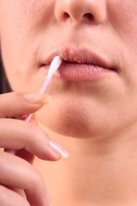
Oral herpes outbreaks are also known as cold sores. An outbreak consists of one or more blisters forming on the chin, cheek, mouth, lips, nose, or throat. Most commonly, the outbreaks occur on and around the lip area. Sometimes canker sores are mistaken for oral herpes. When a herpes outbreak takes place inside the mouth, it is usually is on the top of mouth roof.

Canker sores form in the mouth’s soft tissue areas such as the inside of the cheek. Without proper hygiene, oral herpes can spread to the fingers, and this is called herpes Whitlow. Frequent and proper hand washing is the best method of preventing Herpes Whitlow as simple soap and water kill the virus.

An outbreak of genital herpes consists of lesions and groups of cysts around the genital area. Though both men and women can get genital herpes, it is more common in women, affecting about one out of four.

To date, no cure exists for either oral herpes or genital herpes. Vaccine development is ongoing, and currently the best herpes remedies involve management control with suppressive antiviral therapy. A weak immune system can lead to more frequent outbreaks.

Causes of Herpes

As stated before, contracting either oral or genital herpes is caused by the HSV-1 and HSV-2 viruses. HSV-1 is more commonly associated with oral herpes whereas HSV-2 ir more commonly associated with genital herpes, though either virus can cause either type of herpes.

Herpes is transmitted primarily from person to person through touch. The herpes virus can be present in saliva, flaking skin, and various bodily secretions and fluids such as vaginal discharge and seminal fluid.

A person suffering from either oral or genital herpes does not have to be exhibiting symptoms to transfer the virus to someone else. Some people describe an itching or tingling sensation on the skin before a herpes outbreak occurs. These sensations are a signal that the virus is present on the skin and are termed prodromal symptoms.

A person knowing he or she is experiencing prodromal symptoms can just as easily spread herpes to someone else as a person experiencing the actual outbreaks.

A person suffering from herpes can also not have any symptoms what so ever—prodromal or actual outbreaks—and can still spread the virus. This is called asymptomatic transmission. Some people may have symptoms so mild that they do not even make the connection between their slight itching and herpes. The street name for those that spread the virus via asymptomatic transmission are is Shedders.

Anecdotal evidence also suggests that one can contract herpes from unclean public toilet seats, though no actual hard science backs this theory.

Herpes Diagnosis

Herpes both oral and genital types can be simply diagnosed with a visual exam from a qualified medical doctor.

Usually, with genital herpes a visual exam is only the start of a complete and accurate diagnosis. Several other conditions can bear a resemblance to genital herpes.

Fungal infection or mycosis sometimes can resemble genital herpes. Lichen planus is a reoccurring disease, much like herpes. It causes similar herpes look-alike lesions and rashes. Atopic dermatitis causes the skin to act oddly to normal environs such as food or certain types of clothing. The diseased area becomes itch, flaky, and red and often times is mistaken for genital herpes. Urethritis is an inflammation of the duct where urine is discharged. Urethritis causes difficulties in urinating and pain while urinating. Although urethritis bares no resemblance to genital herpes in symptoms, it is often mistaken by the layperson because of the area in which urethritis effects.

In a case of genital herpes, after a doctor makes a visual examination, the next step laboratory analysis. Samples or a biopsy of the infected area can be tested, as well as blood samples taken. For women, genital herpes can sometimes only occur within the vagina, and so therefore are no outward visible signs of the virus. PAP smears work best in this particular situation.

Preventing Herpes

In the case of oral herpes, do not kiss anyone exhibiting symptoms. If the oral herpes sufferer is experiencing prodromal symptoms, then that person should refrain from kissing others as well to prevent the spread of oral herpes.
Preventing Herpes
It is important to note that oral herpes has the potential to become genital herpes, and the spread of oral to genital can usually be easily prevented. Simply put, if experiencing prodromal symptoms or a full-on oral herpes outbreak, do not participate in oral sex.

As for genital herpes, in general, avoid sex during a outbreak. In between outbreaks, utilize condoms, foams, and spermicides.

Lab tests have shown that the HSV-1 and the HSV-2 viruses can not penetrate latex. Condoms made of polyurethane materials should be avoided because they tend to break, tear, and rip much more often than latex condoms. The use of latex condoms is not a guarantee against the herpes viruses. The latex condom still does break, and a herpes outbreak does not have to be confined to only the genital areas covered by a condom.

Foams and spermicides that contain chemicals such as nonoxynol-9 have been shown in clinical lab tests only to kill the herpes virus. Foams and spermicides should not be used as a replacement of condoms but in conjunction with condoms.

Condoms in conjunction with foams and spermicides, informing the sexual partner about the herpes condition, and avoiding sexual activity during an outbreak can reduce the chances of receiving the HSV-1 or the HSV-2 virus up to forty percent.

Abstinence or complete monogamy with a herpes free partner reduces the risk of herpes exposure to absolute zero percent.

Herpes Risk Factors

Although there is presently no absolute cure for herpes, finding an actual cure seems to be on a low priority list for biological researchers, scientists, and non-profit organizations in comparison to diseases such as diabetes and cancer. This is probably the case because herpes poses no real medical emergency. The virus is painful, annoying, and in the case of genital herpes specifically can sometimes cause depression in a person due to the sensitivity of the area the virus affects.

However, neonatal herpe (herpes contracted by a baby through the birthing process of a mother infected with herpes) can cause mental retardation, severe neurological damage, and even death. If infected with either of the herpes viruses in the genital area, it is critical for the mother-to-be to inform her doctor of the condition.

Neonatal herpes is an exceedingly rare condition though. Out of four million births a year in the United States, rough estimates count neonatal herpes at between 1000 and 3000 cases per year. Out of the twenty to twenty-five percent of pregnant women suffering from genital herpes, less than 0.1% of their newborns contract neonatal herpes.

Still, with human life hanging in the balance, neonatal herpes is a legitimate concern. It’s important to note that neonatal transmission rates are higher for women who contract herpes while pregnant. If the partner has herpes, and the woman does not, it is probably best to avoid sexual relations until the pregnancy has come to full term. About fifty percent of babies that contract neonatal herpes and are treated with the proper antiviral medication do not avoid permanent, lasting damage.

Treatments of Herpes

For treatment of oral herpes, not much can be done except to wait for the outbreak to be over. Management of pain and symptoms seems to be the order of the day.

  • A cold wet towel helps to reduce the swelling of a cold sore if used a minimum of three times a day, and for twenty minutes during each application.
  • Ibuprofen, acetaminophen, and aspirin for those over twenty reduces the pain associated with cold sores.
  • Baking soda mouth rinses can help reduce the pain as well.
  • Avoid eating tomatoes, oranges and other fruits and vegetables that contain high concentrations of acid.
  • Over the counter ointments such as Zilactin, Ziladent or Abreva actually do increase the amount of time it takes to heal a cold sore, and studies have shown can actually prevent the cold sore if applied at the time of prodromal symptoms.

For treatment of genital herpes, not much can done either except to wait for the outbreak to be over. Management of pain, symptoms, and the number of outbreaks one receives per year seems to be the order of the day.

HSV-1 and HSV-2 are direct attacks on the body’s immune system, and outbreaks tend to increase when the body’s defenses are not up to par, so the absolute best method of dealing with herpes is supporting the body’s immune system.

  • Eat right and exercise. Do not over-do the snacks to the point where one becomes overweight. An overweight body tends to reduce the efficiency of the immune system.
  • Decrease the intake of foods with arginine. Arginine is a naturally occurring amino acid that is needed for the herpes virus to replicate. Arginine rich foods include wheat, oats, chocolate, nuts, and gelatin.
  • Increase the intake of foods with lysine. Lysine is a naturally occurring amino acide that seems to block the body’s production of arginine. Foods rich in lysine include fish, chicken, vegetables, and dairy products.
  • Avoid smoking, coffee and other caffeinated products such as soda pop and tea. Avoid alcohol as well, as all of these toxins lead to a less effective immune system.
  • Get enough sleep. Reduce life stresses and worry: these can all trigger an outbreak.

Additional Treatment Options

There are antiviral treatments on the market that help with the pain and sometimes quicken the length of time an outbreak lasts. Prescription drugs are the most common, such as Famvir, Valtrex, and Acydovir. Some people opt for ointments like Dynamiclear. It’s best to see a medical doctor to determine the exact treatment suited specifically to the patient’s individual needs.