Search results for

Dry skin

Conditions

Body Odor

Body odor

Definition

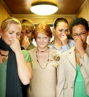
Body odor is a very common problem that everybody has heard of or at least had the chance to smell! Most people understand that body odor and sweat are linked together but they do not know exactly how. According to dictionary.com, body odor is defined as an unpleasant odor from a perspiring or unclean person. There are many different names for body odor, including:

  • B.O.
  • Molodorousness
  • Osmidrosis
  • Bromhidrosis
  • Ozochrotia
  • Fetid sweat

Symptoms & Causes

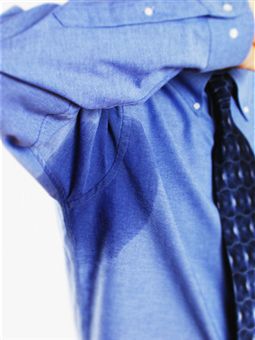
Sweating and body odor are inexplicably linked to one another. Body odor is most likely to be caused by sweat, and sweating is a key reason for body odor. A few things that trigger sweating are exercise, a hot environment or hot weather, and nervousness. These are things that are very normal to most people and so they should be familiar with them.
body odor remedies
Exercise involves exerting the body and causes the body to sweat in an effort to stay cool. The more the body undertakes strenuous activity, the more sweat that is produced.

If the weather outside is hot, those who have to be in the hot area will sweat. Exercise in a hot environment can produce even more sweat than normal.

Anxiety and nervousness are also major reasons for sweating, but cause sweating in specific places such as the face and the palms of the hands.

Sweat Effects

Sweat in and of itself has nearly no smell. It is very faint and can change depending on a person’s mood, diet, hormone levels, or drugs and medical treatments. What this leaves most people asking is this: If sweat is nearly odorless to human beings, why do sweating people always smell so bad?

The answer is that while sweat has no smell, as soon as it is released it comes into contact with bacteria on the skin. These bacteria begin to multiply quite rapidly in the presence of sweat and they cause the smell as well as explaining why it gets worse over time if not properly washed away.

This is especially true of sweat that is the result of nervousness, anxiety, or stress. The body contains two types of sweat glands: eccrine glands and apocrine glands. Eccrine glands are the usual type and are distributed over the entire body. Apocrine glands tend to occur in places where hair grows such as the scalp, groin, and armpits. Eccrine glands secrete sweat when the body temperature rises and their job is to regulate body temperature.

Eccrine gland sweat is composed of water and salt for the most part and does not have an odor detectable to most humans. However, apocrine glands have no role in regulating temperature and instead control pheromones. They secrete a sweat that is fatty and gets pushed directly up onto the surface of the skin. Bacteria then begin to break it down, and the bacteria multiply. It is the breakdown of the bacteria that causes an odor, and this is what is most commonly referred to as body odor.

Risk Factors

Those most at risk for body odor are thus those who are more prone to sweating than others may be. Many studies show that the type of body odor a person has and how it is contrived is genetic. Major histocompatibility complex (MHC) molecules are what largely influence a person’s specific body odor, and they are 100% genetically determined.

How often a person sweats can also be largely influenced by the role of genetics. One study finds that East Asians sweat far less than most others, perhaps in adaptation to their colder climates. Though the studies show interesting results for different groups, one thing is clear for everybody: sweating is a genetic trait passed down and some people have the misfortune of inheriting genes which cause them to sweat heavily in noticeable areas such as the palms of the hands and soles of the feet.

It is also relevant to note that body odor is purely an adolescent and adult problem. Bromhidrosis (an alternative name for body odor) does not occur in humans until they hit puberty, so children do not ever have to worry about body odor.

This can cause confusion in teens who are experiencing puberty and do not realize that they are now at risk for body odor. Body odor is a very easily handled problem once these teens do realize that they must deal with it, but many cultures frown upon unpleasant body odor and so it may be quite embarrassing for them when they do realize this.

When to See A Doctor

Because the tendency of a person to sweat is a genetic trait, it is nearly impossible for doctors to define a “normal” sweating range to use for determining what is abnormal and medically problematic. For this reason, it is important for individuals to know what is normal for them.

Others may judge the amount one person sweats as irregular based on the amount that they sweat, but this would be an uneducated judgment. Instead, individuals should keep in mind what their normal amount of sweating and body odor is and use this to decide if there are any unusual changes or abnormalities occurring.

A change in the amount of sweating or in body odors may signal a problem, and so a doctor’s appointment should be made when this happens. Sweating more or less than the usual amount or randomly having night sweats are both causes for concern.

Body odor changing can be a sign of medical conditions such as diabetic ketoacidosis or kidney failure. Those who sweat excessively may also be interested in seeking medical attention for their problem, as sweating often and in large amounts can be extremely inconvenient and even embarrassing.

Preparing for the Appointment

An appointment can be made with the regular family physician or primary care doctor, or with a dermatologist. It is a good idea to call the primary physician first, as s/he will usually have a good idea of the individual background and might be able to pinpoint things that may be causing the abnormal sweating more easily than a dermatologist who does not have a personal history with a person.

Sometimes the primary physician will immediately refer skin questions to a dermatologist anyway, but it is still good to get a good recommendation from a trusted doctor, and the primary physician can provide this.

Playing the Waiting Game

Most doctors cannot see patients immediately and set up appointments, and once the appointment is set, it would seem that there is not much to do except wait. However, the staff at the Mayo Clinic came up with a list of a few things that people should consider doing while they are waiting for their appointment dates.

The first is to realize that appointments are often brief and time is limited, so a list of questions should be made to ensure that the patient does not forget any important questions that s/he feels need to be asked.

Remember that the doctor does not know everything about the problem the way that an individual does and can only take the information given to try and come up with a solution.

In order to help the process, the patient can write down the symptoms s/he is experiencing, when they first began, and any developments that occurred. Things that seem to worsen the symptoms or help them should be noted on the list as well as how frequently the symptoms occurred.

Tests & Diagnosis

Upon arrival at the doctor’s or dermatologist’s office for the appointment, the physician will usually conduct a physical exam and inquire about the patient’s medical history.
preventing body odor
There are a few simple causes of a change in the amount of sweating or body odor. They are the presence of an overactive thyroid also known as hyperthyroidism or low blood sugar commonly referred to as hypoglycemia. Simple blood or urine tests can determine whether these are the cause of the symptoms and separate courses of action can be taken if they are.

Treatment & Dugs

For most people, a doctor’s appointment will never be necessary in order to solve the problem of body odor. The very simple solutions of over the counter deodorants work for most. Deodorants do not do anything to stop the bacterial breakdown that is the cause of the body odor, nor do they stop the body from sweating.

They are alcohol-based products which help to control the odor by masking it with a more pleasant smell.

For a person whose average day does not involve much physical exertion or one who does not sweat often throughout the day, deodorants regularly applied to the underarms and possibly the hands and feet will be the only solution needed.

Though deodorants solve the problems of those with light sweating, many people exercise on a regular basis or just sweat more throughout the day, and deodorant is not enough to keep them from experiencing unpleasant body odor (and sharing it with everybody around them!).

For these people there are antiperspirants available that will help control body odor in a different way. Antiperspirants do exactly what they say; they help stop perspiration.

Aluminum Compounds

Most antiperspirants contain aluminum-based compounds that block the pores that secrete sweat for as long as the antiperspirant stays on the skin. In this way, less sweat reaches the skin and thus fewer odors is produced. Most antiperspirants contain a deodorant as well to help mask any odor that may still persist.

There are several special brands of deodorants and antiperspirants that strive to offer advanced protection against sweat and body odor. Secret and Gillette are two popular brand names that offer such products. As a personal aside, if you are looking for one such product that will help control body odor and sweating, I highly recommend that you should go online and see how actual users of the products have rated them. There are so many products available that it seems it would be quite difficult to choose one without research!

Most people put on a deodorant or antiperspirant at the beginning of each day and do not worry about the problem of body odor because this solves it effectively for them. For others, more is needed to help control sweating and body odor, and a doctor can suggest and prescribe these special treatments. The most commonly used is an aluminum chloride based antiperspirant such as Drysol or Xerac.

Usage of Antiperspirants

The antiperspirant is usually applied to sweat-prone areas before bedtime and then washed off completely in the morning to help control sweating throughout the day. A normal deodorant and antiperspirant combination usually costs no more than $5 for a stick that will last a few weeks or more.

Special aluminum chloride antiperspirants will cost $17-$25 comparatively and can be bought at drug stores and even Wal-Marts in some places, making them more costly but still relatively quite affordable and easily attainable for the average person.

Risks associated with treatment

Though the risks associated with most normal forms of treatment are small, they should not be ignored. Larger problems than excessive sweating or unpleasant body odor can arise from such ignorance. One very common problem with antiperspirants is that they do their job and block sweat, but nobody really thinks of where this sweat is going. Put simply, think of it this way: The body does not perspire to embarrass people or cause odors. Most of the time it does so because it needs to in order to regulate temperature and it is a natural occurrence.

Antiperspirants block sweat from coming through pores which stops sweat and body odor but can cause a number of other problems. The ingredients of antiperspirants can cause skin irritation which usually manifests itself as a slight burning or stinging that goes away. With the clinical-strength antiperspirants that are obtained from a doctor, this skin irritation tends to be much worse.

Skin Problems

The skin can become red, swollen, and itchy where the antiperspirant is applied. In addition to this, the sweat that was excreted from glands but not allowed to flow onto the skin because of antiperspirant-blocked pores can be trapped and cause cysts and infections. These are all very common problems associated with usage of antiperspirant products to control body odor and sweating.

Very few people have problems with deodorants and thus it is a safer solution, if less effective. Those who do experience skin irritation due to deodorants can usually solve their problems by selecting another deodorant that uses a different ingredient than the one that irritated their skin.

Alternative Treatments & Methods

Hyperhidrosis is the medical term that refers to excessive sweating. Excessive sweating is often the cause of excessive body odor because the two are inexplicably linked together; if the sweating can be solved, the body odor is then also solved. There are many herbal substances that are said to be effective in treating hyperhidrosis and the solutions to it range from these naturally-occurring herbal substances to botunlinum toxin (Botox) injections and surgery.

One such example of a natural herbal treatment is the administration of several herbs in tea form. There are various products available to those familiar with searching the Internet and purchasing products online. One such tea is called Hushed Tea and claims to be able to solve the problem of excessive perspiration through use of such herbs as valarian root, St. John’s wort, and sage. Sipping tea twice a day is much less invasive than botox injections or surgery and is also much more cost effective, and this may be a direction that many sufferers of hyperhidrosis can try for themselves.

Botox Injections

If natural remedies do not work, the next step on the ladder is botox injections. These are quite invasive and can be both painful and costly. Botox injections must be used in all of the places where excessive sweating occurs and the results do not last for very long, typically anywhere from four months to a year at maximum.

Several injections are usually required to obtain the desired results. Even with all of these downfalls, many people turn to botox injections to help deal with their hyperhidrosis because they are actually effective. The botox does not actually solve the problem though; it simply blocks the nerves that trigger the sweat glands and is a temporary fix.

Surgery Options for Body Odor

The final alternative for sufferers of hyperhidrosis is surgery to either remove the sweat glands causing the problems or interrupt the nerve signals to these sweat glands permanently. The Mayo Clinic is a well-known place to have the procedure done and its staff is very experienced with handling this type of surgery.

The success rates for surgery recipients are relatively high depending upon the problem area. For those with overly-sweaty palms, the chances of success are 95-98%. The armpits area has a 75-80% chance of success, and the feet show about a 25% chance of an improvement. Surgery of this type is never done solely to solve the problem of hyperhidrosis if only the feet are affected.

Those who undergo surgery to solve area-specific sweating problems will often notice more sweating on the torso area than they had before as a compensatory means to make up for the sweat that is not being excreted through the former problem area.

Most say that this is not a major problem for them as the compensatory sweat is not nearly as much as was excreted in the original problem area. The only other major difference is the realization that without sweat glands, areas such as the hands can actually become too dry and will need better care.

Prevention

In all reality, there is not much that anybody can do to prevent the fact that all people sweat. Some individuals may do so more than others. However, everybody does it. For most, sweating and body odor are very small problems if they are problems at all and they can be easily solved using the techniques listed above.

For a smaller percentage of people, hyperhidrosis is a problem that causes much stress and anxiety as well as accompanying body odors that can have a substantial effect on the lives of the sufferers. Sweating is a natural occurrence that causes body odor for most and bigger problems for some.

Vitamins

D2

D2 (Ergosterol, Ergocalciferol)

Vitamin D is present in numerous sources of food, such as cod liver oils, fish, eggs and fortified milk. The sun is also responsible for contributing a significant level of vitamin D to a daily production, and it is believed that obtaining just ten minutes of sun exposure is enough to prevent a person from developing deficiencies in vitamin D.

The term “vitamin D” does not technically refer to a single type of vitamin, but rather, it refers to several different kinds. Two forms in particular are of the most importance with regards to human: vitamin D2 (ergocalciferol) and vitamin D2 (cholecalciferol). The former kind of vitamin D is synthesized by plants, whereas the latter kind of vitamin D is synthesized by humans upon the exposure of ultraviolet-B rays directly from the sunlight onto the skin. Some foods may be fortified with both kinds of vitamin D.
d2 deficiencies
The primary biological function that vitamin D has is to make sure the blood have normal levels of phosphorus and calcium in it. It is also capable of aiding with the absorption of calcium, which allows bones to remain strong and for the strong bones to form in the first place.

In recent times, it has been suggested by research that vitamin D may prove to be beneficial in protecting people from several autoimmune diseases, cancer, high blood pressure and osteoporosis.

Two Known Cases

Osteromalacia and rickets are the two classic cases of deficiency of vitamin D. When children experience a deficiency of vitamin D, they will typically develop rickets, the end result of which is a deformity in the skeletal system.

Adults who allow themselves to become deficient in vitamin D will likely develop osteomalacia, the end result of which is weakness in the muscles and the bones becoming weaker than normal.

Those who are at a high risk for developing deficiencies of vitamin D are those who are elderly, are an obese individual, are infants who are exclusively breastfed, and are regularly limited from sun exposure.

Other people at risk are those who have cystic fibrosis, or any other malabsorption syndrome or those who have an inflammatory bowel disease, including Crohn’s disease.

Synonyms

Vitamin D is known by many names, including:

  • 1 alpha D3
  • 19-nor-1
  • 1 alpha-hydroxyvitamin D2
  • 1,25-DHCC
  • 1,25-dihydroxy-22-ovavitamin D3
  • 1,25-dihydroxycholecalciferol
  • 1,25-dihydroxyvitamin D3
  • 1,25-diOHC
  • 1,25 2D3
  • 1-alpha-hydroxycholecalciferol
  • 22-oxacalcitriol
  • 25-dihydroxyvitamin D2
  • 25-dihydroxyvitamin D2
  • 25-HCC
  • 25-hydroxycholecalciferol
  • 25-hydroxyvitamin D3
  • 25-OHCC
  • 25-OHD3
  • activated 7-dehydrocholesterol
  • activated ergosterol
  • alfacalcidol, calcifediol
  • calcipotriene
  • calcipotriol
  • calcitriol
  • cholecalciferol
  • colecalciferol
  • dichysterol
  • dihydrotachysterol 2
  • dihydrotachysterol
  • ecocalcidiol
  • ED-21
  • ED-71
  • ergocalciferol
  • ergocalciferolum
  • hexafluoro-1
  • 25dihydroxyvitamin D3
  • irradiated ergosterol
  • MC903
  • paracalcin
  • paricalcitol
  • viosterol
  • vitamin D2 and
  • vitamin D3

Evidence

Familial Hypophosphatemia

Familiar hypohosphatemia, which is the instance of the blood having low levels of phosphate, is a disorder that is both rare and inherited, which consists of the transport of phosphate being impaired in the blood, as well as the vitamin D metabolism present within the kidneys being diminished. Familial hypophosphatemia is a known kind of rickets. Familial hypophosphatemia suffers are able to take phosphate supplements, in addition to dihydrotachysterol or calcitriol in order to effectively treat some of their bone disorders. However, management should be made beneath the supervision of a physician.

Fanconi syndrome related hypophosphatemia

Fanconi syndrome, associated with renal tubular acidosis, is a defect associated with the proximal tubules within the kidney. Taking oral dosages of ergocalciferol has been found to be useful towards treating instances of hyophosphatemia that is associated with the presence of Fanconi syndrome.

Hyperparathyroidism Associated With Low Levels of Vitamin D

Some patients who have low levels of the essential vitamin D may find themselves with secondary hyperparathyroidism. As people may expect, the necessary and initial treatment for resolving this issue is to supplement a person with vitamin D. It is commonly recommended to have surgical removal of the parathyroid glands in those who have either refractory or primary hyperparathyroidism, however. There are some studies which suggest the supplementing a person with vitamin D should reduce the occurrence of hypoparathyroidism that may follow an instance of surgery in order to treat primary hyperparathyroidism, whether the parathyroid glands were partially or wholly removed.

Hypocalcemia Resulting From Hypoparathyroidism

It is rare to experience hypoparathyroidism, or having low levels of the parathyroid hormone in the blood, and it usually only occurs after removing the parathyroid glands through surgery. Taking in large doses of calcitriol, dihyrotachysterol or ergocalciferol may help with increasing concentrations of serum calcium in people who suffer from pseudohypoparathyroidism and hypoparathyroidism.

Osteomalactia, or Adult Rickets

Adults that allow their levels of vitamin D to sink far enough below to undergo severe deficiency turn out to develop osteomalacia, the loss of mineral content in the bones, the weakness of bone muscle and pain in the bones. Elderly people who do not have enough vitamin D in their diets, those who do not get an adequate amount of sun exposure, those who have an inability to absorb vitamin D, those with kidney disease and renal osteodystrophy, those who have had intestinal or gastric and those who have had a bone disease due to aluminum are all at risk of developing osteomalacia. The underlying cause of the osteomalacia ultimately determines what the best course of treatment is. For the most part, it involves taking phosphate binding agents, vitamin D, orthopedic surgical intervention and controlling the pain.

Psoriasis

There are a few different ways to treat the different forms of psoriasis skin plaques. A couple of the more mild approaches might include stress reduction, light therapy, moisturizers or even salicylic acid in order to remove the areas where scaly skin develops. In cases that are a little more severe, it may be necessary to include treatments of retinoids, such as isotretinoin (Accutane), UV-A light, corticosteroids, psoralen plus UV-A light or cyclosporine. Vitamin D3 appears to be able to control the growth of skin cells and is thus used when skin plaques are moderately severe.

Uses Based Upon Theory and Tradition

The uses as described below are based upon studies and tradition. Because some of these conditions may be very serious, it is important to first consult a doctor prior to taking any vitamin D for treatment.

  • Actinic keratosis;
  • Alzheimer’s disease that results in fractures of the hip;
  • Ankylosing spondylitis;
  • Autoimmune disorders;
  • Graves disease;
  • Hyperparathyroidism, particularly in renal dialysis;
  • Hypocalcemia;
  • Hypocalcemic tetany;
  • Kidney transplant causing bone loss;
  • Metabolic disorders;
  • Nervous system disorders, such as hemichorea;
  • Osteitis fibrosa through dialysis;
  • Rheumatoid arthritis;
  • Scleroderma;
  • Squamous cell carcinoma;
  • Systemic lupus erthematosus;
  • Vaginal disorders; and
  • Vitiligo.

Dosages

Adults (over 18 years old)

Most multivitamins have vitamin D included in them, and it is usually in strengths ranging from 50 IU to 1,000 IU when they are in tablets, softgels, liquids and capsules. The following levels of AI, or Adequate Intake, have been established herein by the United States Institute of Medicine.

It is recommended for all individuals, whether male, female, lactating or pregnant, to take in 5 micrograms of vitamin D on a daily basis, which also amounts of 200 IU. This only applies to those under the age of 50 years. Individuals who are between the ages of 50 years and 70 years should consider taking 10 micrograms on a daily basis, which also amounts to 400 IU. Those who are over the age of 70 years are recommended to take 15 micrograms on a daily basis, which also amounts to 600 IU per day.

There have been some authors who questioned the validity of these results, stating uncertainly with whether or not it is sufficient to meet with a person’s physiological needs, especially if they are an individual that does not regularly expose themselves to the sun adequately. The upper limit for obtaining vitamin D has been recommended to be 2,000 IU on a daily basis, based on reported toxicities with higher dosages.

Not all listed doses have previously been proven to be affective for certain conditions. However, in an oral dosage of 400 – 800 IU per day, ergocalciferol has previously been used to treat and prevent osteoporosis.

Children (under 18 years old)

It is recommended for newborns and children of all ages to take only five micrograms on a daily basis, which amounts to 200 IU per day. Children who are older than a year should not take more than the upper limit for their age, which is 50 micrograms on a daily basis, which amounts to 2,000 IU per day. Children who are below the age of one year are recommended to not exceed the maximum limit of 25 micrograms on a daily basis, which amounts to 1,000 IU per day.
d2 uses
It is potentially unsafe to take vitamin D in excess amounts orally, with potentially advertise affects such as high levels of calcium in the blood, or hypercalcemia.

There have been some authors who questioned the validity of these results, stating uncertainly with whether or not it is sufficient to meet with a person’s physiological needs, especially if they are an individual that does not regularly expose themselves to the sun adequately.

A review from 2008 has recommended that all infants and children, as well as all adolescents, should only take 400 IU on a daily basis based on evidence resulting from historical precedence and current, new clinical trials.

Safety

The United States Federal Drug and Food Administration is not known to strictly regulate the usage of herbs or supplements. There is no way to guarantee any strength or purity based on the usage thereof. Prior to beginning any new therapies, it is recommended to consult a physician.

Allergies

Anybody who is known to have a hypersensitivity to vitamin D or its derivatives or analogues should avoid taking it.

Side Effects and Warnings

With the recommended AI dosages, vitamin D is mostly well tolerated, though a study found that patients who took vitamin D analogues did experience a greater likelihood in experiencing daytime sleepiness.

It is possible to experience toxicity when regularly taking excess dosages of vitamin D. The end result may include excess bone loss or hypercalcemia. Those who have kidney disease, tuberculosis, sarcoidosis, hyperparathyroidism or histoplasmosis are at especially high risk. Pre-existing instances of chronic hypercalcemia should be managed by a doctor due to its potential to lead to serious or life threatening complications.

Early symptoms of this condition may include anorexia, nausea, vomiting, excess thirst, excess urination, fatigue, weakness, somnolence, dry mouth, headache, tinnitus, vertigo and ataxia. Function of the kidneys may become impaired and there may be an instance of calcium being deposited into several organs, which will also affect the kidneys. The necessary treatment for these is to stop taking calcium or vitamin D and to lower the levels of calcium under only the strictest medical supervision. It may be necessary to use corticosteroids and to acidify urine.

Pregnancy and Breastfeeding

It is recommended for pregnant women to take the same level of vitamin D as if they were not pregnant. There have been some authors who felt that pregnant women should take in more vitamin D, especially if sun deprived, but this has yet to be clearly established. Discuss higher doses of vitamin D with a physician due to the risk of vitamin D toxicity.

Maternal milk does not have much vitamin D to it, so it may be necessary to supplement an exclusively breastfed infant for the first two months of life in order to prevent vitamin D deficiency.

Vitamins

Tocopherol

Vitamin E – Tocopherol (Alpha, Beta, Gamma, Delta)

Tocopherols are a group of four chemical compounds discovered in 1922 by anatomist and embryologist Herbert McLean Evans. Evans was conducting feeding experiments on rats using vitamins B and C. During these experiments, Evans concluded that despite the rats being feed every known nutrient, they were still infertile.

Upon feeding them wheat germ, the rats were able to become pregnant. After years of experiments and studies attempting to isolate the unknown compound in wheat germ that allowed the rats to become fertile, Evans found the formula C29H50O2 and concluded that it reacted like an alcohol, making it part of a hydroxyl group. Evans named the compound after the Greek words “tokos” (birth) and “pherein” (to bear or carry) meaning, “to bear young”, adding the –ol to indicate the alcohol property of the compound. The structure of Tocopherols was later determined in 1938, over twenty years after the initial experiments.

Types

Tocopherols are a lipid-soluble (fat-soluble) antioxidant that, when combined with corresponding tocotrienols, make up the common dietary supplement Vitamin E. However, almost all Tocopherols and tocotrienols have Vitamin E activity and properties, and can be correctly referred to as Vitamin E on their own. The four forms of Tocopherols are alpha, beta, gamma and delta.

Alpha

Tocopherol is the form of vitamin E most easily absorbed by the human body, and found in the largest quantities in human blood serum. It is believed to be responsible for protecting cell membranes and reducing the risk of cardiovascular problems. It is the most found tocopherol in vitamin E supplements and in European diets.

Beta

Beta-tocopherol has lower vitamin E activity than the other three tocopherols and amounts found in human blood serum are lower than with alpha, gamma and delta tocopherols. While technically providing the same benefits of the other three tocopherols, it is not as easily absorbed by the human body and therefore is used only in small amounts in vitamin E supplements.

Gamma

Tocopherol is second only to alpha-tocopherol in its vitamin E activity, and is the most common tocopherol found in the American diet. While it is higher in concentration than alpha-tocopherol in food sources, the liver removes it from human blood serum, causing it to be found in lesser amounts in the human body than alpha-tocopherol. Because of this, it is not an effective lipid antioxidant.

Delta

Tocopherol has a lower concentration in food sources than alpha-tocopherol or gamma-tocopherol, yet is more easily absorbed by the human body than gamma-tocopherol. Therefore, it is primarily used with alpha-tocopherol in vitamin E supplements, and it is believed that when delta tocopherol is combined with other tocopherols in a vitamin E supplement, the supplement is more easily absorbed by the human body.

Sources and Dosage

Doctors recommend that a 25-year-old male ingest 15 milligrams a day of vitamin E. Due to the fact that high amounts of vitamin E can act as an anticoagulant (preventing the clotting of blood), it is recommended that vitamin E intake not exceed 1,000 milligrams per day. Two American surveys in 1991 and 1994 indicated that most Americans do not consume the recommended amount of vitamin E, though a study by the Institute of Medicine in 2000 suggested that the surveys could be flawed because added fat during cooking is often under-reported. They believe that most Americans actually do receive the recommended amount of vitamin E from their normal diets, though caution those who consume low-fat diets to take a vitamin E supplement due to the lack of vegetable oils, a main source of vitamin E.

Besides vegetable oils, vitamin E is also found in high concentrations in nuts, seeds and whole grains. However, most Americans receive the bulk of their vitamin E from breakfast cereal and tomato sauce. Vitamin E is found in these sources, listed from highest to lowest: Wheat germ oil, sunflower oil, almond oil, sunflower seeds, almonds, hazelnuts, walnut oil, peanut oil, olive oil, poppy seed oil, peanuts, pollard, maize, poppy seeds, asparagus, oats, chestnuts, coconuts, tomatoes, walnuts, carrots and goat’s milk. While wheat germ oil contains 215.4 milligrams of vitamin E per 100 grams, goat’s milk only has .1 milligrams of vitamin E per 100 grams.

Synthetic Types & Supplements

Synthetic vitamin E is also added to packaged foods, especially cereals. Most fortified breakfast cereals can contain upwards of 24 milligrams of vitamin E per serving, which is well above the recommended intake of 15 milligrams per day.

For those consuming a low fat diet, vitamin E supplements are recommended. Natural supplements usually derive vitamin E from soybean oil, while synthetic supplements usually use straight alpha-tocopherols or a combination of alpha-tocopherols and one or two of the other tocopherols. Natural vitamin E supplements, while often more expensive, are recommended over synthetics as they are absorbed by the human body in larger quantities and more efficiently than synthetic vitamin E. Some supplements contain both natural and synthetic tocopherols, and are usually priced between the all natural and all synthetic versions of vitamin E.

Medical Usage and Health Benefits

The proper intake of vitamin E can help prevent and theoretically treat a host of common health concerns.

Alzheimer’s disease, a progressive form of dementia, is believed to be partially caused by oxidative stress. Studies suggest that high intake of vitamin E can prevent cell membranes from oxidizing, and therefore can be used as both a preventative measure and treatment for Alzheimer’s disease. However, studies are mixed as to whether vitamin E actually helps, with some trials indicating it does and others that it does not. Nonetheless, vitamin E has not been found to speed up the progression or onset of this disease.

Antioxidants such as vitamin E have been proven to help prevent the effect of free radicals on the human body, which are known to contribute to the development of chronic diseases such as cancer. Studies indicate that vitamin E may block carcinogens, a known cancer causing substance, from accumulating in the body, thereby preventing the development of certain cancers.

Vitamin E is also known to boost immune function, protecting those already with cancer from additional threats to their already weakened immune system. Most studies suggest that vitamin E primarily protects against prostate cancer and breast cancer, though it has not been shown to decrease the risk of breast cancer in postmenopausal women. On the contrary, recent studies have indicated that high intake of vitamin E by smokers can actually increase the risk of lung cancer by seven percent for each 100 IU of vitamin E taken daily.

Protect Your Eyes

Vitamin E has been found to protect the human eye from a host of diseases. When combined with other antioxidants, has been found to slow the onset and progression of age-related macular degeneration (AMD). AMD is an eye disease that usually develops after the age of 60 and the leading cause of vision problems and blindness among the elderly. However, vitamin E by itself has not been found to have any effect on the development of AMD.

Observational studies have indicated that regular use of vitamin E supplements in those that already consume the recommended amount of vitamin E in their diets reduces the risk of developing cataracts. In 2007, a study found that when vitamin E supplementation was added to other treatments for glaucoma, a disease damaging the optic nerve of the eye, it helped protect the spread of glaucomatous damage to the retina. Those treated with additional vitamin E showed significantly less vision impairment than those who were not.

Preventing Parkinson’s

In 2005, a study was published suggesting that vitamin E could help prevent the onset of Parkinson’s disease, a degeneration of the central nervous system that progressively impairs motor skills and speech. People who consumed foods high in vitamin E were found to have a lower risk of developing this devastating disease, though consuming these foods after the onset of Parkinson’s disease has not been shown to have any effect on the progression of the disease. In addition, use of supplemental vitamin E had no effect on either the onset of the disease or the progression.

Preventing Heart Disease

Vitamin E is widely believed to prevent or delay coronary heart disease, though this has only been found in small studies and disputed in larger studies. Vitamin E has been shown to reduce LDL-cholesterol (also known as “bad” cholesterol) and the formation of blood clots. LDL-cholesterol increases the risk of blockages in coronary arteries, which leads to atherosclerosis and heart attacks. Observational studies have indicated that those with a higher than usual intake of vitamin E were thirty to forty percent less likely to develop heart disease.

Despite this, clinical trials do not show the same outcome, though research is ongoing. Some studies indicate alpha-tocopherol only vitamin E supplements actually increase the risk of heart failure. When alpha-tocopherol was combined with gamma-tocopherol, this was not the case. The most promising use of vitamin E in regards to heart problems was found by the American Heart Association in 2007, when they stated that women taking regular doses of a vitamin E supplement were 21% less likely to suffer a blood clot than those who were not, though continuation of blood thinners was recommended for treatment.

Other Uses

Tocopherols are often used as a preservative in foods to prevent oils from going rancid, especially in dry pet food. A mix of all four tocopherols (alpha, beta, gamma and delta) has been found to be most effective for this application. Vitamin E is also widely used in cosmetics and as an additive in packaged foods. It is believed to be good for the skin, and is found in most moisturizers and foundations. Its predominance in these products, while possibly beneficial, is primarily because vitamin E is inexpensive. While marketing often preaches the benefits of vitamin E when applied topically, the research behind this claim is shady at best. Recent studies have indicated that 90% of study participants noticed no improvements in their appearance when vitamin E was added to a product and a third of these actually developed rashes from synthetic versions of vitamin E.

While vitamin E, when consumed according to guidelines, can be beneficial to one’s overall health, it can also increase morbidity rates when taken in excess. As with all things regarding one’s health, it is best to discuss vitamin E supplementation with one’s doctor.

Sources:

Conditions

Respiratory Diseases

Respiratory Diseases are ailments and conditions that impair our respiratory system. The respiratory system is responsible for proper delivery of oxygen throughout the body, removal of carbon dioxide and toxic waste, ample regulation of the body temperature and the stabilization of the body’s pH level. Respiratory diseases are those that affect the lung, bronchial tubes, pleural cavity, trachea, upper respiratory tract and all muscles and nerves involved in the breathing process.

There are many reasons why respiratory diseases occur. They could result from an infection, accident or from inhaling some toxic products. It could also be genetic or a byproduct of unhealthy habits such as smoking. Genetics also play a part in contributing to the occurrence of respiratory diseases.

Types of Respiratory Diseases

There are several types of respiratory diseases. Let us briefly define them and look at some examples of each type.
respiratory disease symptoms
Chronic Obstructive Pulmonary Disease (COPD)

Lung diseases that result from a severe obstruction of airflow Mto the lung are termed as a chronic obstructive pulmonary disease or COPD. These diseases would damage and narrow the airway and block airflow in the lungs.E.g.: Emphysema

Obstructive Lung Diseases

This is a form of lung disease that occurs when the airflow in the bronchial tubes get narrowed, restricting airflow to the lungs.

E.g.: Asthma, Silicosis

Restrictive Lung Diseases

Lung diseases of this form occur where there is a reduction in lung volume either because they cannot stretch enough to alter its volume according to the pressure applied, or due to diseases affecting the pleura or chest wall.

E.g.: asbestosis, Sarcoidosis and pulmonary fibrosis

Respiratory Tract Infections

Upper respiratory tract infection

Essentially, these are infections that occur to the upper portion of the respiratory system. Some examples are common cold, sinusitis, tonsillitis, otitis media, pharyngitis and laryngitis.

Lower respiratory tract infection

The most common example of an infection affecting the lower portion of the respiratory tract is pneumonia.

Respiratory Tumors

Respiratory tumors are either malignant or benign. While the benign ones such as pulmonary hamartoma rarely cause respiratory diseases, lung cancer caused by malignant tumors pose a significant health problem. Lung cancers accounts for 15% of all cancer reports and 29% of all deaths due to cancer.

Pleural Cavity Diseases

Pleural cavity diseases occur when fluid gathers in the pleural cavity during pleural effusion or when a hole in the pleura, called pneumothorax causes the affected lung to collapse.

E.g.: Emphysema and Mesothelioma

Pulmonary Vascular Diseases

Diseases are regarded as pulmonary vascular diseases when they impact the pulmonary circulatory system of our body. They could occur from a pulmonary embolism, pulmonary arterial hypertension, pulmonary edema and damage to the capillaries in the lung that results in blood leaking into the alveoli.

Symptoms

Respiratory diseases can be detected with some of these many symptoms. One should definitely seek medical consultation and help if they find themselves suffering from one or more of these symptoms.

  1. Shortness of Breath with or without exertion
  2. Breathing Difficulties or Dyspnea
  3. Rapid Breathing
  4. Dizziness and Fainting
  5. Chest Pains
  6. Accelerated Heart Rate of more than 100 beats per minute. This is also known as Tachycardia.
  7. Fatigue
  8. Loss of Appetite
  9. Weight Loss
  10. General uneasy feeling
  11. Bluish discoloration of the lips, tongue and/or fingers, also known as cyanosis.

Risk Factors

There are many factors that could contribute to the increase in occurrence of respiratory diseases. Constant exposure to these factors could increase the chances of respiratory diseases occurring.

  1. Tobacco smoke
  2. Indoor air pollutants such as those that could come from open stoves, mold, dust mites, and pollen.
  3. Outdoor air pollutants that can arise from exhaust fumes from cars and factories, smoke, dust and smog.
  4. Allergens that can comes from pets, pollen, dust mites, mold, smoke, formaldehyde and volatile organic Compounds (VOC).
  5. Occupational hazards such as exposure to toxic fumes, chemicals and harmful substances
  6. Unhealthy Lifestyle such as smoking.

Tests and Diagnosis

To determine if one is suffering from any type of respiratory disease and to determine which type, several tests could be ordered by the physician. The type of test that would be administered on a patient depends on their medical history, family history, symptoms and the diagnostic procedure physicians are expected to follow.

Some of the tests are

  1. 1. Chest X-Ray
  2. Chest X-ray is a radiograph projection of the chest that helps physicians analyze and diagnose respiratory diseases. It uses ionizing radiation to create images of the chest. X-rays are used to determine conditions that involve the chest wall, thorax bones, lungs and heart. The occurrence of pneumonia is commonly determined through an X-ray.

  3. Pulmonary Function Tests (PFTs)
  4. The most common PFT that is used to measure lung function is spirometry or the measurement of breaths. This is a tool that is used to create pneumotachographs that can be used to determine respiratory conditions like asthma, pulmonary fibrosis, cystic fibrosis and chronic obstructive pulmonary disease (COPD).

  5. Computed Tomography Scan
  6. Also known as a CT scan, this is a medical imaging procedure that uses tomography to create a three-dimensional image of the body part. High resolution computed tomography scan is used to diagnose respiratory diseases like emphysema and fibrosis. A general purpose image reconstruction technique is sufficient to check for pneumonia and cancer.

  7. Examination of microorganism cultures derived from secretions like sputum
  8. A microbiological culture is created when microbial organisms are allowed to multiply in a controlled environment and then tested to determine the cause of an infectious disease. In some cases, a chemical test is conducted on the culture to determine and eliminate certain causes of infection or diagnosis and could be vital information to determine the form of treatment that needs to be administered to the patient.

  9. Bronchoscopy
  10. This technique allows the insides of the airway to be viewed. Bronchoscopy also helps to provide therapeutic relief. The bronchoscope is inserted into the airway through the nose, mouth or a tracheostomy. It helps physicians analyze the airways for abnormalities like blood, tumors and inflammations.

  11. Biopsy of the lung or pleura
  12. This medical test involves the examination of cells or tissues that are removed from the patient. These are examined through a microscope by a pathologist and could be chemically analyzed.

  13. Ventilation/Perfusion Lung scan (V/Q lung scan)
  14. This form of medical imaging that uses scintigraphy to assess the blood and air circulation within the lungs. The ventilation part of the scan allows on to determine how air reaches all parts of the lung and the perfusion part assesses how blood circulates within the lungs.

  15. Ultrasound
  16. Ultrasounds are especially useful to determine the effect of pleural effusion. Pleural effusion occurs when excess fluid gathers in the pleural cavity of the ling. This could limit the expansion of the lungs and affect their breathing ability. The four types of fluid that could accumulate and cause pleural effusion are serous fluid, blood, chyle and pus.

    These tests would help determine the exact type of respiratory disease that has occurred and would allowed doctors to determine the type of treatment that needs to be administered.

Treatment Options

Many factors affect the type of treatment that would be administered for respiratory diseases. These factors are the type of respiratory disease, its intensity and the medical history of the patient in concern. Here are some possible treatment options that would be suggested to help manage one’s respiratory disease.

  1. Medication
  2. Patients could be prescribed with corticosteroids, bronchodilators, antibiotics, anticoagulants, cancer chemotherapy and immune suppressants to help them with their respiratory diseases.

    Side effects of these medications vary from one medicine to another. All patients must ensure that doctors know their full medical and family history to determine which form of medication is the safest and would be most suitable for them. Patients would also be warned of any specific side effect that could occur from the consumption of the medication and they should understand them clearly and comply as needed. Cost of these medications would vary based on the location, availability of medical facilities and how the insurance of the patient covers the medicinal cost.

  3. Physiotherapy
  4. Physiotherapy is most used when one has had a pulmonary surgery or has been diagnosed with cystic fibrosis, COPD or pulmonary fibrosis. Physiotherapy helps clear lung secretions and certainly help those who have suffered from respiratory diseases.
    herbal respiratory disease remedies

  5. Oxygen Therapy
  6. This is helpful when one is unable to inhale in sufficient oxygen due to the respiratory disease that their body is suffering from.

    This form of treatment is primarily administered when patients suffer from emphysema, pneumonia and any respiratory disorder that impacts the oxygen intake of the body.

    This form of treatment is flexible and can be administered in the hospital, at home and could be made portable as well.

  7. Mechanical Ventilation
  8. This process involves the mechanical replacement of the natural breathing process with ventilators. This method involved a ventilator and a trained individual who compresses the bag as required. This could be used as a short term measure during an operation or for chronic illnesses as well. Possible side effects are complications such as pneumothorax, injury of the airways and alveola, and ventilator associated pneumonia.

  9. Radiotherapy
  10. This process utilizes high energy rays to kill some or all cancer cells. In early stages of cancer, it could completely remove the cancer. In other cases, it can be supplemented with chemotherapy or surgery. In more advanced lung cancer, radiotherapy treatments could keep the illness under control by shrinking the cancer cells, providing some symptomatic relief for the patient.

  11. Surgery
  12. Surgical procedures like lobectomy and pneumonectomy could be performed to remove the cancer. Pleurodesis is a procedure that artificially eliminates the pleural space. Lung Volume Reduction Surgery where the damaged portions of the lung could help with COPD and emphysema patients. A patient’s damaged lung could be partially or completely replaced through lung transplantation. Surgery also allows for an artificial lung to be implanted into the body.

Home Remedies to Treat Respiratory Diseases

While there are many treatment options, there are some natural home remedies that one could take to control their respiratory diseases. These natural remedies could be consumed to provide relief from the symptoms of the respiratory diseases, cure the diseases to some extent and keep them in control. Needless to say, these natural remedies do not replace the doctor’s advice and prescription. When one intends to undergo a strict natural regimen, they should consult their doctor and do it as a supplement to the medications and treatments that is being administered to them.

Ginger

Ginger is a perennial herb that belongs to the rhizome family. The medicinal values possessed by gingers have been recognized for a long time by ancient Indians, Chinese and Greek physicians. Ginger is also used as a spice in many cultural dishes. The vitamins and minerals found in ginger are phosphorus, calcium, iron, carotene, thiamine, vitamin C, niacin and riboflavin. Ginger provides a great remedy for coughs and colds. When consumed with honey, ginger could provide relief from dry cough. Ginger tea is also a great remedy for colds and fever.

Fever resulting from influenza could be reduced from a mixture consisting of ginger juice, fenugreek and honey. It also works as an expectorant in asthma, bronchitis, whooping cough and tuberculosis.

Chicory

Chicory is a perennial herb that has long tap roots, round stems and several light or dark green leaves and pale blue flowers. This herb is native to the Mediterranean region and was known to the ancient Greeks and Romans. Its ability to create better skin was discovered a long time ago. Chicory is helpful for asthma and hay fever recovery. Juices of chicory, carrot and celery after eliminating starches and sugars help with these respiratory conditions. Powder derived from the dried chicory roots mixed in honey is a great remedy and expectorant for chronic bronchitis.

Celery

This salad plant consists of green leaves, stem and bulbous roots. The celery roots and seeds are popular in Unani and Ayurvedic treatment and medicines. The Chinese and English have used celery as a medicine since the fifth century B.C. Celery contains a well balanced proportion of nutrients, vitamins and minerals. Its antispasmodic properties help in treatment of respiratory diseases like asthma, bronchitis, tuberculosis and pleurisy.

Tumeric

Tumeric is a perennial herb that has short stems and raised branches. This rhizome has been used by those who practice Unani and Ayurvedic medicine since ancient times. The antiseptic properties of turmeric make it a good remedy to treat chronic cough and infected throats.

When fresh turmeric powder is mixed in warm milk, they make a good remedy concoction that would help increase the discharge from the nose and provide relief from cough and cold. It is also used as a great in house remedy for bronchial asthma.

Prevention Tips

One could prevent the occurrence of respiratory diseases by adhering to at least some of the prevention tips that are mentioned here. While these prevention tips may not entirely eliminate the chances of suffering from a respiratory disease, they would at least reduce the chances of respiratory diseases from occurring. It could also help with the healing process and ensure that the infectious respiratory diseases do not spread across the population. These measures are also good for general health of all within the family and society.

  1. Do not smoke. If you are, work on quitting soon. Smoking would damage your lungs and the lungs of those surrounding you, especially close family and friends. Smoking is the primary cause for many respiratory diseases and you and your loved ones are succumbed to.
  2. Educate the young children about the hazards of smoking. It would be better to educate them at an early age to ensure that they can stay protected from this unhealthy habit and protect their lung health.
  3. Avoid inhaling second-hand smoke. Being a passive smoker by inhaling smoke also affects your lungs and increases your chances of being affected by a respiratory disease.
  4. Be more proactive and protect your family from passive smoking. You could divert from smokers who are in your way. You could also instruct visitors to your home that smoking would not be tolerated.
  5. Always be clean and thoroughly wash your hands. Fight germs by maintaining your hygiene. The spread of infection reduces when everyone does their part to remain clean and germ free.
  6. Cover your coughs and sneezes to prevent the spread of flu, cold and other lung diseases.
  7. Contribute to limiting outdoor air pollution. Do your part to ensure that everyone breathes in cleaner air.
  8. Find out how you could reduce the amount of indoor air pollutants within your house.
  9. Ensure that you exercise regularly to build stronger lungs and immune system.
  10. Avoid occupational hazards such as inhaling toxic fumes from chemicals, paints and other solvents. If needed wear a protective mask and ensure that good air quality is maintained at your work place at all times.
  11. Ensure that you have taken the necessary vaccinations to protect yourself from respiratory diseases that are prevalent in your area.

Conclusion

Respiratory diseases come in many forms and severities. Learning more about them, how they occur, how they can be treated and prevented would certainly help us manage our lung health better. This is vital information whether you or a loved one is currently managing a respiratory disease.

Conditions

Bedwetting

Bedwetting is commonly defined as involuntary urination during sleep in children after the age that bladder control normally occurs. It can happen any time the child is asleep, whether day or night, but most commonly occurs during nighttime sleep. Nocturnal enuresis is the term used to describe instances of involuntary urination at night.

Primary nocturnal enuresis is the condition in which a child has not yet achieved an extended period without wetting the bed. Secondary nocturnal enuresis is diagnosed when an individual, either a child or adult, returns to wetting the bed again after having been dry for some time. Although thought of as a childhood condition, many adults suffer from nocturnal enuresis as well, estimated at around 26 million adults in the U.S. according to the Simon Foundation for Continence.

Symptoms

The primary, and often only, symptom of nocturnal enuresis is the involuntary release of urine during nighttime sleep. Other symptoms may occasionally be present, such as daytime wetting, general incontinence, increased urgency to urinate, increased frequency of urination, burning during urination, and constipation.

Causes

Nighttime wetting has numerous potential causes, with the enuresis likely influenced by more than one of the following. Most cases of bedwetting fall under the category of primary nocturnal enuresis. PNE cases are occasionally caused by medical conditions, but most can be attributed to a developmental issue. In these cases, the body has not yet matured enough to stop urine production at night. This results in the bladder filling up during a period where awareness of the need to urinate is diminished due to sleep.
Bedwetting Cures
The body naturally develops the ability to delay urine production during sleep by around age 5. Genetics can also play a role, as children whose parents were not bedwetters have only a 15% chance of becoming bedwetters themselves. In contrast, children with one or two parents who wet the bed exhibit a 44% and 77% chance of developing enuresis respectively.

There are other less common causes of bedwetting, and since there are no clear tests to determine if enuresis is caused by a developmental delay or genetics, the following more-easily diagnosed causes are often either identified or ruled out first.

Medical Conditions

Several medical conditions can cause a person to wet the bed. Infections and certain illnesses, such as a urinary tract infection, can insight involuntary nighttime urination, though the likelihood of this is estimated at less than 5% of all cases. Physical abnormalities might also be a cause of bedwetting, primarily due to a smaller than normal bladder.

In children, a condition in which an insufficient amount of the anti-diuretic hormone (ADH) is produced might also play a role. This hormone normally increases at night, signaling the kidneys to slow urine production. If low levels of ADH continue throughout the night, the bladder will continue to fill often leading to a wet bed.

Psychological Issues

It is not uncommon for bedwetting to result from psychological trauma or anxiety. Mental or emotional issues resulting from a death in the family, negative social pressures, physical or sexual abuse, or a hostile home environment, can manifest themselves through nighttime wetting. Enuresis itself is not regarded as a psychological disorder, but it can be symptomatic of a troubled psyche.

Sleep Disorders

Certain sleep disorders have also been identified as contributing to instances of bedwetting. Sleep apnea, a condition where the airway becomes obstructed during sleep, can be a potential cause of nocturnal enuresis. Sleep walking can also be a cause as the sleep walker often dreams they are in another room of the house, and thinking they are in the bathroom, they urinate only to find upon waking that they have wet the bed.

Diuretics

Substances which increase urine production, such as caffeine, dark chocolate, and alcohol, are called diuretics. These substances work opposite any ADH signals active in the body, telling the body instead to produce more urine rather than less.

Other Causes

A few other potential triggers thought to contribute to enuresis include the following. Stress is an often documented cause of a return to bedwetting, or secondary enuresis. The constant tension in the body experienced by those under heavy stress wears out the muscles, and leads to difficulty maintaining stamina in the sphincters associated with the bladder at night. Heavy sleeping can also lead to bedwetting. Though children have been shown to wet during all five sleep stages, parents who report that their children are heavy sleepers also report higher rates of bedwetting.

This could be due to the difficulty for those children to transition from the sleep state to being awake, causing difficulty in resuming bladder control once the ADH hormone begins to diminish. Constipation can also be a cause as the excess fecal matter can press against the bladder making the need to urinate more urgent. Some believe food allergies might also be potential cause of enuresis, though more documentation is needed. One controversial cause of nighttime accidents is aggressive or improper potty training. Training too early or with too much negative reenforcement has been sighted as a possible environmental cause of bedwetting, due to the anxiety children might experience with such training.

Diagnosis

With bedwetting, diagnosis usually begins with an in-depth consultation between the family physician and the parent or guardian of the child. In some cases, the child may be asked some questions directly. These questions might include how many times has the child wet the bed, have they ever been dry, is there any family history of bedwetting, and are they taking any medications currently. He might be curious about the diet of the child, or whether the child has ever wet during the day.

Other questions will also be asked specifically to rule out some of the less-ambiguous potential causes.Following the consultation, and based on the answers given, a series of tests will be run to attempt to diagnose the cause of the wetting issue or to rule out even more potential causes, allowing the doctor to narrow down the list of possible triggers.

The physician will likely take a urine sample, and X-rays may be necessary if the doctor feels as though a physical abnormality might be behind the enuresis. With secondary enuresis, especially if the individual has reached middle age or beyond, a more extensive evaluation might be given, taking into account more psychological factors. In many cases however, it proves easier to pin-point the cause of secondary enuresis, since it often corresponds with an event, injury, or treatment.

Treatment

Treatment for bedwetting varies from medical intervention to counseling. It is very specific to the identified cause of the enuresis. If a physical abnormality is identified as the source of the issue or if the bladder was damaged in an accident, surgery may be performed to strengthen the bladder walls or repair the urinary tract. If a medical condition, such as an infection or Diabetes, seems to be the cause, medications may be prescribed. In the majority of cases, neither surgery nor medication end up as the recommended treatment for nighttime wetting.

The most often recommended treatment in cases of primary nocturnal enuresis is simply to wait for the child’s body to mature, since most physicians do not expect a child to be accident-free until age 5 or so. Another common treatment prescribed for bedwetting is the use of a bedwetting alarm. These alarms sound when they sense wetness in the crotch area, causing the child to wake and become aware of the sensation of a full bladder. They have proven to be considerably effective as over time, the child begins to awaken from sleep on their own when they experience the now-familiar bladder pressure.

Synthetic hormones, such as Desmopressine, may be prescribed in cases where the anti-diuretic hormone is not being produced in sufficient quantities. If the cause seems to be psychological in nature, anti-depressants may be prescribed.

Punishment, as a tool to stop bedwetting, has been proven ineffective in several major studies on the subject, and has been shown to increase the frequency of bedwetting rather than reduce it.

Coping

Bedwetting can be traumatic for a child, causing embarrassment, social awkwardness, and the feeling that something is wrong with them. For adults or children who return to wetting, it can cause frustration at the inability to control their own body, and depression due to the struggle to regain continence, which is often dealt with in secret. For many individuals, treatment does not bring with it an instant cure, and bedwetters must deal with wet beds, stained pajamas, and heavy hearts in the mean time.

Several coping methods exist to deal with wetting the bed. The first, and most obvious, is diapers or absorbent briefs. Heavy wetters will likely need an overnight diaper, such as Depends, to prevent urine from reaching the bedding and soiling pajamas. The average bedwetter however, can usually opt instead for absorbant underwear, such as Goodnights or Pull-Ups. These briefs are less bulky than a standard diaper, and pull-on like underwear.

Product Design

They are increasingly being made to look like underwear, to the point of felling like cloth, and even mimicking the waist-band. There are absorbent underwear options for the child, all the way up to the youth and adult age groups. These options make it less embarrassing for a child to attend a sleepover or camp-out, and therefore less psychologically draining. It should be noted that some believe that excessive use of protective underwear can actually prolong bedwetting, since the individual cannot feel the resulting wet bed as they normally would. Another coping option is the use of a mattress cover.

These are employed to prevent the mattress from becoming stained with urine, and the accompanying smell. Finally, almost all bedwetters are advised to limit the intake of fluids several hours before bed. This gives the bladder time to produce the remaining urine from the days meals, which can be voided before bed, leaving the body less likely to produce a significant amount of urine over night.

Prevention

A consistent sleep schedule, allowing the individual to get a full-nights rest, and regimented visits to the bathroom go a long way in preventing accidents at night. Also, avoiding enuretic triggers, such as diuretics, before bed should also be observed.

Complications

There are few complications that arise from wetting the bed, but the following should be considered. Rashes and other skin issues may result from sleeping in wet clothing over an extended period. Diaper rash cream or petroleum jelly can ease the discomfort and prevent further inflammation. As mentioned before, emotional or psychological issues may result from bedwetting, and should not be dismissed as insignificant.

Alternative Medicine

Several alternative therapies have been used to treat enuresis, and these treatments have been gaining popularity. Hypnosis has been used to varied success on individuals who wet at night, focusing on suggestions to stay dry or to wake to use the toilet upon certain triggers. Acupuncture has also been used to treat bedwetting, with some success. There are several homeopathic remedies for nighttime wetting, but consider before use that these have been determined to be ineffective in clinical trials.

Conditions

Fever

Fever, also known as pyrexia, is any body temperature that is elevated above the normal temperature of 98.6 degrees Fahrenheit. Body temperature fluctuates throughout the day, and can elevate due to normal everyday activities, such as exercise. However, an elevated temperature of 100.4 degrees Fahrenheit or higher is considered medically significant, and is usually due to an underlying medical condition.

How to Diagnose a Fever

If the body temperature is higher than the normal temperature of 98.6F, and cannot be explained by activity level, a fever is present. Diagnosing this is a simple matter of taking one’s temperature with a thermometer. Sometimes, depending on signs and symptoms accompanying the fever, a physical exam or other tests will be ran in order to determine if the cause of the fever is infection or something else. Once a determination of the underlying medical condition has been made, other tests (such as a blood test) may be required to confirm the diagnosis.
Fever Symptoms

How to Determine Body Temperature

A thermometer is used to measure body temperature. The three most common methods are rectal; oral; tympanic (in the ear); and axillary (under the armpit). Rectal is most often used with infants and is the most accurate method. Oral is the next most accurate method, and is also the most widely used in children above the age of 4, and adults. The tympanic method requires a digital thermometer, and is often used in children over the age of 3. Axillary is the least accurate method.

Taking Temperatures

Rectal Method (infants)

  • Use small amount of lubricant on end of rectal thermometer
  • Place infant on belly and carefully separate buttocks cheeks
  • Carefully insult the thermometer ½ – 1 inch into the infant’s rectum
  • Keep hand cupped loosely on infant’s bottom and keep fingers on the thermometer so the thermometer and baby are still
  • Wait for the thermometer to beep, or wait for three minutes if not using a digital thermometer

Oral Method

  • Clean the tip of the thermometer with soap and warm water or rubbing alcohol. Rinse with cool water
  • Put tip under tongue
  • Close lips gently around thermometer
  • Keep thermometer under tongue until thermometer beeps, or for three minutes if not using a digital thermometer

Tympanic Method

  • Put a new, throw-away cover over the tip of the thermometer
  • Keep head still
  • For child, gently pull child’s ear straight back; for adult, gently pull ear up and then back
  • Put covered thermometer tip into the ear opening. DO NOT push hard.
  • Press the button to turn on the thermometer and hold the button until the thermometer beeps (or follow manufacturer instructions)
  • Remove thermometer from the ear opening

Axillary Method

  • Place thermometer in the armpit with arms crossed over chest
  • Wait for the beep, or wait four to five minutes if not using a digital thermometer

Causes of Fever

Our body temperature is set by the hypothalamus, which is an area in the brain that acts as a thermostat for our bodies. The thermoregulatory set point is determined by balancing the heat produced by body tissues such as the liver and muscles, and the heat lost by the body.

The thermoregulatory set point is increased in response to threats, such as bacterial or viral infections, resulting in a higher body temperature as the body directs blood away from the skin to decrease heat loss. When the fever occurs, there are feelings of chilliness, stiffness and shivering caused by the body generating heat until the blood around the hypothalamus reaches the new, elevated set point. When the body temperature then begins to return to normal, there is often sweating as the body attempts to get rid of the excess heat.

The most common cause of a fever is infections, such as the flu or a cold. However, other possibilities include

  • inflammatory conditions such as rheumatoid arthritis
  • extreme sunburn
  • heat exhaustion
  • surgery
  • heart attack
  • hemorrhage
  • hyperthyroidism
  • lupus
  • inflammatory bowel disease
  • malignant tumor or some form of kidney cancer
  • side effect of some immunizations
  • side effect of some medications

Symptoms of Fever

  • dehydration
  • general weakness
  • headache
  • inability to concentrate
  • loss of appetite
  • muscle aches
  • sweating
  • trembling, shivering

High fevers between 103F and 106F may cause

  • confusion
  • convulsions
  • hallucinations
  • irritability

Febrile Seizures

Children under the age of 5 can experience fever induced seizures, otherwise known as febrile seizures. A febrile seizure occurs when a child’s temperature rises or falls quickly. The signs of a febrile seizure include convulsions and a brief loss of consciousness.

Febrile seizures occur in approximately 3% of children between the ages of 18 months and 3 years. Of those children, 1/3 will have another seizure with febrile episode. These seizures do not generally cause long term side effects or damage to the nervous system

When to Seek Medical Attention

Low fevers are generally not cause for alarm. In fact, if the fever does not cause discomfort and there are no troubling symptoms accompanying a low fever, it should go untreated to allow the body’s natural defenses to combat the underlying illness. However, there are times when medical attention is necessary.

Medical attention should be sought when

  • temperature is 104F or higher (seek IMMEDIATE medical attention! A temperature this high can lead to delirium and convulsions)
  • a child under 3 months of age has a temperature of 101F or higher
  • infant refuses to eat or drink
  • fever in infant is accompanied by unexplained irritability, or unusual crying during a diaper change or when moved
  • fever in infant is accompanied by lethargy and unresponsiveness
  • fever in a child occurs after being left in a hot car
  • there is a history of cancer, HIV, AIDS, heart disease, diabetes, or other serious illness
  • other symptoms suggest major illness
  • fever is sustained for more than 3 days

Risk Factors

As fevers are usually signs of an underlying medical condition, the risk factors for a fever go hand in hand with the risk factors for the underlying medical condition. Exposure to illness, family history of more serious ailments causing fever, and a weakened immune system are all risk factors for contracting a illness, and thus for experiencing a fever. The exact factors putting individuals at risk for developing a fever depend greatly on the cause of the underlying medical condition that causes the fever.

How to Prevent a Fever

In order to prevent a fever, exposure to underlying medical conditions must be reduced. The most effective way to reduce exposure to the underlying medical conditions causing fever is to wash hands frequently, particularly before eating, after using the restroom, after exposure to a crowd or someone who is sick, and after petting animals.
Fever Prevention
Developing good hand washing practices is imperative to prevention, and should be taught to children as soon as possible.

Avoiding touching the “T-zone” of your face—eyes, nose, mouth—helps to prevent fever by reducing the spreading of viral infections.

In addition to hand washing and avoiding touching the “T-zone” of your face, covering your mouth when coughing and nose when sneezing helps to prevent the transmission of infection, and thus prevents fever.

Three rules to prevention

  1. Wash hands frequently
  2. Cover your cough and sneeze
  3. Avoid the “T-zone”

Treatment

High-grade temperatures, as opposed to low-grade, may be treated to lower their severity. The treatment for fever depends greatly on the underlying medical condition causing the fever. If the underlying medical condition is a bacterial infection, prescription medications such as antibiotics will be prescribed.

However, antibiotics are ineffective against viral infections. For any underlying illness causing the fever that cannot be treated with prescription medications, over-the-counter medications or home remedies may be used. It is important to note that these over-the-counter medications and home remedies do not cure the fever or the underlying medical ailment. Rather, these treatments alleviate the symptoms of the fever.

Over-the-counter medications

Over-the-counter medications used to treat a fever include acetaminophen (Tylenol), ibuprofen (Advil or Motrin), and aspirin. These help to lower a high fever, but will not cure it. Acetaminophen should not be used if the person with a fever has liver disease. Adults may take 2 extra strength tablets (totaling 1000 mg) every 4-6 hours.

Children should seek medical advice. Ibuprofen is safe for children over 6 months in age, though medical advice should be sought regarding proper dosage. Adults may take 2-3 200mg tablets every six hours. Aspirin should NOT be used with children or adolescents, due to concerns with a serious illness called Reyes Syndrome. Aspirin is, however, safe for adults.

Home Remedies

There are various home remedies that, while they will not cure the underlying illness, will help the fever run its course, and make a person suffering from a fever feel better

  • Stay hydrated. Drink water, juice, or broth. Fever causes fluid loss and dehydration so it is important to keep sipping something in order to keep the body hydrated.
  • Rest. Activity can raise body temperature, so plenty of rest is recommended to help body temperature return to normal
  • Soak in lukewarm water. Tepid water (85F) baths may help bring temperature down. Soak for 5-10 minutes for a cooling effect. Do not use alcohol, and if the bath causes shivering, stop the bath. Shivering raises the body’s temperature, so will have an opposite effect on the fever.
  • Herbal Tea. Various recipes of tea are available that claim to aid in the reduction of a fever. These include Basil Leaf Tea drank 3-4 times per day during the fever; tea with ½ teaspoon cinnamon powder, 2 cardamoms, and 2 teaspoons ginger boiled well; 2-3 teaspoons dry roasted fenugreek seed powder boiled and strained with a drop of butter added; and 10 grams raisins and ginger boiled in 200 ml water until it is reduced to 50 ml, with the water then strained and drank.
  • Herbs. Various herbs, independent of the herbal tea, are thought to be good treatments for fever. 7-8 tulsi leaves (basil) and 3-4 peppercorns chewed together is supposed to help a fever, as is a teaspoon of honey with 2-3 drops of ginger juice.

While high-grade fevers may be treated to alleviate the symptoms of the fever, low-grade fevers should not be treated with either over-the-counter medications or home remedies. Treating low-grade fevers may extend the length of the illness and interfere with the body’s immune response. In these cases, allowing the body to combat the illness is the best option.

While fevers are the body’s natural defense against illness, it is important to remember that sometimes our body needs a little help. Don’t put off seeking medical attention if the fever is accompanied by worrisome symptoms, if the fever is exceptionally high, or if the fever does not decrease after taking medication.

Conditions

Piles

Piles, better known as hemorrhoids, are located within the anal canal. There are typically two classes of piles: external and internal. Each indicates the location of the hemorrhoids. Once swollen or inflamed, they become pathological. Piles are one of today’s most common ailments, affecting several people.

Hemorrhoids

Piles, or hemorrhoids, is classed as a varicose and is an inflamed condition of the anal veins, either within or just outside the anus. In the case of external piles, there will be a great deal of pain but not much bleeding present. When internal piles are present, there is a common discharge of a dark blood that is secreted. In the event that the veins burst, there will be a condition called bleeding piles.
Piles
Piles are seen as round swellings that are located within the anal canal, in the area termed as the anal cushions. Since there are many people that never seek treatment or consult a physician for piles, it isn’t known just how many cases there are. There are several different types of piles categorized in different degrees, based on symptoms and severity.

  • First degree: Swellings located on inside lining of anal canal. Occasional bleeding but aren’t seen on the outside.
  • Second degree: Large and protrude from anus during bowel movement, returning afterwards.
  • Third degree: Same as second degree however do not return until pushed in.
  • Fourth degree: Hang down permanently, not able to be pushed back inside the anus. Blood within can become clotted causing extreme swelling and pain.
  • External: Swollen veins that develops below the anal cushions and are typically more painful than other types.
  • Sentinel: Lumps that develops from anal fissure that heals and creates a painless skin tag.

There are other types as well, presenting different characteristics and locations. The pain typically increases towards the higher degrees and the more outer anal cushion piles.

Initial Symptoms & Causes

The symptoms can vary depending on the type, as well as the treatment. Fiber, oral fluids, NSAIDs, rest, and sitz bathing has been shown to be the most commonly recommended treatment. Rest is especially important as there are many complications associated with overexertion and overstress.

There are surgical measures that are taken when the piles does not respond to other treatment options. Many home remedies and herbal remedies are available, as well as over-the-counter medications that can be applied. A physician can also prescribe various medications to aid in the relief of the symptoms and the treatment of the piles for more severe cases.

There are several causes of piles, typically dependent upon the type and severity. Many of the causes are associated with functions of the anal canal, typically from some sort of irritation or strain that causes the veins to enlarge and many times protrude. Typically any over exerted activity can risk the development of piles, and should be avoided. Though some cases bleed, there are some that do not. However, treatment should be sought out to avoid progression of the affliction for any case. Worsening will continue through time causing them to get larger and more severe, especially if there is no change in stress or inexertion of the area.

Symptoms

There are a great many symptoms associated with Piles, each characteristic of the condition. The most common piles cases can show the least symptoms, sometimes going unnoticed for a bit of time until the inflammation and swelling increases. Many times, these cases will even often times resolve on their own in a few days. Typically, hemorrhoids are benign and are simply enlarged and swollen veins protruding the anal cavity below the anal cushion.

The external piles cases tend to be more painful and itchy, while the internal piles can be painless. Most commonly, the symptom of internal hemorrhoids is noticed during a bowel movement in which the stool will be covered by bright red blood.

This is a condition that is called hematochezia, and can be noticed in the toilet bowl or the toilet paper. Sometimes the internal piles will protrude through the anus, while many times they won’t. External hemorrhoids can be seen as a swollen lump around the anus, which will inflict a great deal of pain when inflamed.

The most common symptoms of hemorrhoids are:

  • Itching of the anus
  • Aching or pain around the anus, especially when sitting
  • Bright red blood in the stool
  • Painful bowel movements
  • One or more painful, hard, and sore lumps around the anus

There are some cases when there can be a slimy mucus discharge of the anus as well, indicating there is a case of piles or even that the bowels haven’t been emptied entirely. These cases are typically dependent on the severity and type of piles that is present.

Causes

The actual cause for piles isn’t completely understood or known. However, there are theories that concern the cause, one being that there is a weakness of the anal tissue connecting the anal cushions too the muscle layers located underneath. When there is a straining during bowel movement, this anal cushions can slide out of place and down into the rectum. This presents itself as one or more hemorrhoids and can also be associated or complicated with diet.

Eating a low fiber diet can assist in causing the piles as the bowels will be much harder and more difficult to pass without strain. This strain passing these bowels could result in one or more piles developing. Constipation for this reason, is another thought to be cause of piles, which highly increases chances of developing the affliction.

Who Piles Effects

Piles are thought to afflict the elderly more so than the younger adults as the rectum structures aren’t supported as well. Pregnancy has a strong influence in the development and many women who are pregnant are at a high risk of developing the hemorrhoids. As the fetus bears down on the lower half of the woman’s body and as she strains to walk comfortably, these piles will erupt in mild to severe cases, sometimes resolving shortly after child birth. There are also some children born with a weak rectum structure, or have a family history of piles, which increases the risk of development.

Other causes are extended periods of sitting or standing, extremely strenuous working conditions, obesity, mental tension, and various bowel disorders.

Risk Factors

There are several risks, most which correlate with the causes. Heredity, age, activity, and diet can play a large role in the risk for piles. Pregnant women are at an extreme risk due to the extra strain that is placed on the anal cushions. With the rapid weight gain and the disproportioned weight in the mid section, there is an increase risk for excessive strain.

The elderly are also at risk as age deteriorates muscle structure and strength. Children are typically not affected by hemorrhoids, but children can be born with weakened muscles in the rectum that will increase their risk of developing piles later in life. Constipation and other bowel disorders have a strong influence on the risk for developing the condition as this creates extreme strain and pressure.

There are 5% of Americans that are afflicted at one point of their lives with piles. Over one million people in the US are afflicted each year with hemorrhoids, yet many others don’t even report the condition, leaving many cases unaccounted for.

Prevention Tips

The best prevention method for piles is to maintain soft stool so there is no strain during bowel movement; this decreases the pressure and strain during bowel movement. It is also a necessary to prevent piles, to pass stool as soon as the urge occurs. Daily exercising, including jogging and walking, as well as a diet rich in fiber can reduce the chances of constipation and will assist in making softer stools so they are easier to pass. There should be minimal time attempting to defecate as this could create the slippage in the anal cushion that develops piles.

Test and Diagnosis Considerations

If there is bleeding occurring from the rectum, a physician should be contacted immediately in order to decrease any complications that may occur from the piles. The physician will typically diagnose according to description of the symptoms as well as an examination of the rectum and the anal canal. The exam will consist of inserting a gloved finger into the rectum gently to feel for any hemorrhoids. There will also likely be an evaluation of medical history.

Physicians also may use a proctoscope for looking inside the anal cavity. This proctoscope is a narrow, tube-like camera that will blow air through to open the bowel and see more clearly. This is a great test for ruling out the occurrence of worse complications that may be causing the bleeding and pain. There is also a sigmoidoscopy or colonoscopy test that is done within the hospital allowing the doctor to see inside the large bowel. These are tests that are typically done in order to ensure there isn’t a more serious condition causing the symptoms.

Treatment Options

Although there are many treatment options for piles that can alleviate the symptoms and provide a great amount of relief from the swelling and pain, there is no cure for the condition. There are several home treatments that are basic and consist of relieving the symptoms through self help methods. There is also a great deal of medical treatments, and there are surgeries that can take place in the most severe cases of piles. Over-the-counter creams are great for treating the symptoms and are most commonly used.

Herbal and Home Remedies

The most common home remedy is to bath in sitz which is an antimicrobial liquid that can be poured into the bath to sooth any inflammations and prevents infections of many kinds. Other herbal and home remedies include the use of:

  • Dry figs
  • Mango seeds
  • Jambul fruit
  • Radish
  • Turnip
  • Bitter Gourd
  • Ginger

There are many other home treatments that include using onion, wheat grass, sesame seeds, and many other types of extracts. The most common remedy however is also a fiber rich diet that allows for softer and more passable stool. Exercise is also quite important to the treatment as it allows the bowels to remain strong and able to pass stool easily. There are also various laxatives that can be used to soften the stool when constipation.

Medical Treatment

There are several over-the-counter corticosteroid creams that are effective in alleviating pain and swelling. Witch hazel applied with a cotton swab can also be effective at reducing itching followed by several other steps including:

  • Wear cotton underwear
  • Perfumed or colored toilet tissue
  • Avoid scratching as it will irritate and inflame more

Increasing fiber intake, oral fluids, and use of NSAIDs and taking advantage of rest can also be very effective at treating the condition, even decreasing the size of the hemorrhoids. Preparation H is a commonly used cream for piles, but will not cure the condition or make it go away. Many times there are flair ups that must be treated, as they occur, which could be often or seldom.

There are also various medical procedures that will be performed without surgical need, including:

  • Rubber band ligation: elastic bands applied to internal hemorrhoid to eliminate blood supply, allowing it to wither and fall off. Cure rate is seen at 87%.
  • Sclerotherapy: injection of sclerosing agent, including phenol, into the piles. The vein walls collapse and the hemorrhoid shrivels up and falls off. The success rate of this cure is 70%.
  • Cautery: many methods such as electrocautery, infrared radiation, and cryosurgery are performed to remove piles.

Surgical Treatment

If there is no response to medical treatment, there are some surgical techniques that are performed. They can cause urinary retention, or other complications associated with the surgery, as the surgery is performed so close to those organs. Types of surgery include:

  • Hemorrhoidectomy: surgical excision of hemorrhoid used in severe cases. Requires 2-4 weeks for recovery.
  • Doppler guided transanal hemorrhoidal dearterialization: taping off of Doppler located arteries providing blood to the piles. Minimal complications yet has high reoccurrence rate.
  • Stapled hemorrhoidectomy: resection of soft tissue, disrupting blood supply to piles. Less painful than removing the hemorrhoids and has faster healing rate.

These surgical treatments are highly effective at removing the hemorrhoids; however there is no guarantee that there won’t be recurrence. The most severe cases are relieved with surgical measures, but these measures are much more costly than other non-surgical treatments.

Conditions

Urticaria

Urticaria is commonly known as hives, uredo or needle rash and is a type of allergic reaction that causes the skin to welt into red blotches. The welts are extremely itchy and can cover large areas of the human body including arms, legs, face, stomach and back. The cause of urticaria can be related to an immune response to food, other allergens or contact with an allergenic substance. Urticaria can also be related to excess stress. Urticaria can also present after a viral infection like mononucleosis, German measles or hepatitis.

Closely related to urticaria is a type of swelling called angioedema, which causes welts that are embedded deeper in the skin, particularly near the lips and eyes. Most of the time urticaria and angioedema will resolve without treatment in a short period of time, but there are cases that can be life threatening, especially with angioedema if the throat and/or tongue swell enough to block the airway. Angioedema and urticaria can occur at the same time and can be severe.
Urticaria Definition
Angioedema can be caused by food allergies, medications like aspirin and latex allergies. Hereditary angioedema is related to a C1 inhibitor deficiency or lack of normal blood protein.

Different Types of Urticaria

There are basically four types of urticaria including acute, chronic, drug induced and physical. Acute urticaria comes on rapidly after touching an allergen. It can last as long as several weeks to as little as a few hours. Allergies to food and certain product ingredients fall into the ‘acute urticaria’ category. Common allergies to food include nuts, eggs, fish, shell fish, food dyes and acid derivatives. Common allergies to products include many different types of ingredients used in perfumes.

Chronic urticaria is a case of hives that lasts six weeks or longer. There are cases reportedly lasting as long as 20 years or more. There is no real known cause of chronic urticaria and it is often referred to as idiopathic in nature because of this.

Drug induced urticaria is serious and can result in cardiorespiratory failure. One of the main drugs known to cause drug induced urticaria is the anti-diabetic suphonylurea glimepiride or Amaryl®. This drug is documented as to causing severe allergic reactions, which appear as urticaria. Penicillin, aspirin, ACE inhibitors, sulfonamides, anticonvulsants and NSAIDs can also bring on or exacerbate angioedema and chronic urticaria.

Depending on cause, physical urticaria can be categorized into several categories including:

  • Cold reactions like cold air, water or ice.
  • Cholinergic reactions to exercise, after a hot shower or body heat
  • Aquagenic reactions to water (extremely rare)
  • Delayed pressure after standing for long periods of time, or to items like belts and bra straps
  • Dermographism from skin scratching
  • Heat reactions to hot objects or hot food
  • Vibration
  • Solar reaction to sunlight
  • Adrenergic reaction to noradrenalin or adrenaline

Risk Factors For Urticaria and Angioedema

The risk factors for urticaria and angioedema increase if a person has experienced either in the past, has allergic reactions, suffers from disorders like lymphoma, thyroid disease or lupus, has a family history of angioedema, hereditary angioedema or hives.

Symptoms of Urticaria

There are several symptoms associated with urticaria that include severe itching, stinging and burning. Hives emerge as elevated, erythematous or papules, plaques and linear streaks that are transient and usually encompassed by a flare or halo. The lesions are usually pale, particularly in the center. They can vary in size from several centimeters to a few millimeters.

Hives brought on by heat, exercise, changes in temperature or emotions and induced by acetylcholine appear as morphologically unique lesions that usually disappear in an hour or less. Lesions caused by trauma or scratching have a streaking linear appearance that is pronounced with a surrounding flare. In angioedema, pale areas of edema with diffused borders can be recognized on various parts of the body and face. If angioedema affects the larynx, dyspnea, stridor or hoarseness can occur and can be quite serious, even leading to death.

In cholinergic urticaria, induced by acetylcholine and triggered by emotions, heat, exercise or changes in temperature, the lesions are morphologically different and disappear in 30-60 minutes. Dermographic lesions, induced by brisk scratching or trauma, are linear streaks with a surrounding flare.

Symptoms of Angioedema

Angioedema, as noted, is quite similar to hives and recognized clinically as pale areas of edema deeper in the skin. Angioedema appears as firm, thick, rather large welts, skin swelling, blistering and pain. Angioedema mainly appears on the eyes and lips but can also present on hands, genitalia, feet, or inner throat. Involvement of the larynx may lead to hoarseness, stridor and dyspnea, and may be fatal. Hereditary angioedema is uncommon and more serious than common urticaria or angioedema. It is identified by rapid and severe swelling of various body parts, as well as abdominal cramping if the digestive tract is involved.

Visiting a Doctor For Urticaria and Angioedema

As mentioned, most hives and angioedema episodes are not life threatening and can be treated at home. But in more chronic and severe cases, an appointment with a doctor to determine the cause and offer treatment is highly recommended. As a general rule of thumb, see a doctor if hives or angioedema does not go away after a day or two or if a home remedy or over the counter treatment (see Treatments Section Below) does not work in relieving symptoms. If symptoms include breathing difficulty, swelling of the throat or fainting and/or a lightheaded feeling, go to an emergency room for immediate treatment.

A doctor will take a patient’s history to try to identify the cause of urticaria or angioedema. It helps if a patient brings notes to the appointment that include important information like signs and symptoms of urticaria or angioedema, how often the symptoms occur, how long they last and any medications, herbs or vitamins taken to relieve symptoms. A patient should also write down any questions he or she might have regarding tests, causes, symptoms, alternate approaches, and best course of action to take.

A doctor will ask questions like:

  • When was the onset of symptoms first noticed?
  • How did the urticaria or angioedema present in appearance?
  • Have symptoms changed at all?
  • What makes symptoms worse?
  • Have any home remedies or over the counter medications been used?
  • Have prescription medications been taken?

The answers to these questions will help the physician put together a complete history and proper diagnosis of the condition and how it might best be treated.

Following a complete history and evaluation, the physician may do a scratch or prick test to help identify any allergies to foods, insects, pollen, animal fur, medications, etc. The doctor may also do a patch test, which is an allergen placed on a patch and applied to the skin. If it causes a reaction, the allergen is identified and the appropriate treatment can be initiated.

Similarly, the doctor may do an intradermal test, whereby an allergen is injected into the skin. Intradermal testing is usually performed if the physician suspects an allergy to insects or penicillin. A doctor suspecting a patient might be suffering from hereditary angioedema, may request a blood test to evaluate levels and blood protein function.

Treatment Options For Urticaria and Angioedema

If treatment for urticaria or angioedema is recommended, standard antihistamines should help. Antihistamines will block the release of histamine, which causes the symptoms and discomfort of urticaria and angioedema. Over the counter medications include:

  • Diphenhydramines like Benadryl
  • Chlorpheniramines like Chlor-Trimeton
  • Loratadine like Claritin and Alavert
  • Cetrizines like Zyrtec

Some of these medications can cause drowsiness, so caution should be taken before medicating. For instance, a patient should not drive when taking antihistamines, unless the medication states that it does not cause drowsiness.

Prescription medications include:

  • Desloratadine or Clarinex
  • Fexofenadine or Allegra
  • Hydroxyzine or Vistaril
  • Levocetrizine or Xyzal

If the hives or angioedema are severe, a physician may prescribe a corticosteroid drug like prednisone to help bring down swelling and ease itching.
Urticaria Medications
To treat hereditary angioedema, a physician may prescribe a long term treatment of androgens like danazol, which help maintain and regulate blood protein levels. There are also many new medications that are in clinical trials that might be recommended.

If the episode of hives or angioedema appears to be severe or life threatening, an emergency adrenaline injection may be necessary. This will require visiting an emergency treatment center immediately. If these severe episodes are frequent, a physician may recommend and prescribe adrenaline that can be used by a patient in an emergency.

After self injecting an adrenaline, a patient should go to an emergency room for further treatment if necessary.

Side Effects of Antihistamines

As mentioned, drowsiness can be a side effect of taking antihistamines for urticaria or angioedema. Other side effects include changes to the immune system. A patient should speak to a physician about side effects and complications of mixing antihistamines with other medications.

Side Effects of Corticosteroids

The side effects of corticosteroids can be serious affecting the joints and other parts of the body. That is why, when prescribed, they are used for a short period of time in hopes of alleviating severe symptoms of urticaria or angioedema. Most corticosteroids must be withdrawn gradually to avoid further complications.

Home Remedies and Lifestyle Changes To Help Relieve Urticaria and Angioedema

Rather than taking over the counter medications or prescribed medications, some patients choose to try home remedies and lifestyle changes to help urticaria and angioedema symptoms. To relieve itching and pain associated with urticaria and angioedema there are natural remedies and lifestyle changes that could help significantly. They include:

  • soaking in oatmeal baths
  • soaking in detoxifying baths
  • rose water and vinegar applications
  • mint and brown sugar drinks
  • Turmeric powder
  • the herb rauwolfia mixed in a cup of water
  • applying anti-pruritic lotions.

Lifestyle changes include switching to an all fruit diet for five days, maintaining a balanced food diet of nuts, seeds, fruits, grains and vegetables and avoiding foods like coffee, tea, alcohol, and foods that are hard to digest.

Epson salts baths in hot water are claimed to be the most effect way to relieve outbreaks of urticaria or angioedema. The patient should soak in a hot bath treated with a cup of Epson salts for 20 minutes or more at least three times a week. Do not use soap as it may introduce harmful chemicals and prevent the positive effects and results of the Epson salts.

There is also a chemical free technique or Organic technique that is proving quite helpful for treating chronic urticaria. The theory is by wearing and using clothes and other items like sheets and bath towels that have not been exposed to laundry detergent chemicals, toxicity levels are greatly reduced in the body, and the immune system is boosted.

After about four to six weeks, urticaria and angioedema could disappear. To successfully carry out this technique, a patient must use chemical free, cold water methods to wash items they come in contact with. High levels of baking soda should be added to the long rinse wash cycle, and all items must be air-dried to prevent heat combustion that can cause chemical residue in a dryer.

Preventing Outbreaks of Urticaria and Angioedema

Active patient participation is important in preventing outbreaks of urticaria and angioedema. There are ways to lower the chances of outbreaks by staying away from known triggers like foods that seem to bring on hives or angioedema, medications or situations that provoke onsets. Situations can include anything from temperature changes, stressful environments and exposure to chemicals that could provoke an allergic reaction.

It is also highly recommended that patients keep a log of their outbreaks of urticaria or angioedema, noting what foods seem to cause reactions, what environment brought on a reaction like extreme heat or cold and what chemical (for instance, a certain component in a perfume) caused an allergic attack. By using these self-help techniques, many times a patient can identify and eliminate the cause or causes of their urticaria or angioedema attacks and avoid taking over the counter or prescription medications and the side effects that go along with them.

Conclusion

It is worth the time invested to research urticaria and angioedema to know the various degrees and forms of both conditions. In most cases, urticaria and angioedema are temporary and not serious, but there are rarer forms of urticaria and angioedema that can present serious and life threatening conditions.

Patients experiencing hives or mild angioedema should benefit by taking an antihistamine that can help relieve swelling and itching. In severe cases, corticosteriods may be prescribed for a short period of time. If a patient experiences any difficulty in breathing, swallowing or maintaining consciousness, immediate emergency treatment must occur. Some patients with severe reactions carry epinephrine injections and antihistamine pills to administer should a severe reaction occur.

Hereditary angioedema is not an allergic reaction. It is a genetic disorder that affects the immune system. It can be triggered by injury, infection or stress and the main symptom is swelling, particularly around the mouth and eyes. If swelling occurs in the windpipes, breathing can be inhibited and immediate attention is required. Certain treatments may relieve hereditary angioedema symptoms.

Conditions

Gray Hair

Gray Hair Facts

Hair follicles, like skin, bear pigmentation in the form of melanin. For hair, there are two types of melanin: eumelanin (brown pigment) and pheomelanin (black pigment).These are the same melanin types that create the classic stripes of a Tiger or spots of a leopard. Their different combinations will result in blond to brown to dark brown, to black hair colors. The balance is similar to paint mixing with only two colors, and results in the many hair color shades in between blond and black, in all populations of the world.

The hair can grow with a number of combinations of these two pigments and their concentrations are biochemically controlled by their stem cell instruction set, DNA.

Melanin Loss

Grayness is directly related to melanin loss or deficiency. The average person has roughly a 3:1 active /recessive hair growth ratio. For every 4 follicles, 1 is dead/dying, 3 are active and growing. This growth is not a regenerative process in terms of the fact that a follicle does not keep making hair after hair during its cellular lifecycle; it is a one-time-build. Each hair has a new follicle manufactured just for that one hair growth cycle.
Gray Hair Facts
When a hair follicle dies the keratinocyte and melanocytes recede and expire in a pre-programmed physiological manner. The hair falls out and the follicle remains are reabsorbed by the body. During this process stem cells (progenitor cells) which are located at the base of the follicle, begin to build new keratinocyte and melanocytes, leading to the growth of a new hair.

Scientific Voids

The graying of hair is a result of a disruption in this process – in particular, lack of melanin production. Free radical damage has been highlighted as a possible cause, but scientists worldwide have yet to pinpoint the exact science behind hair graying – strenuous investigation is ongoing to find the causes (there is definitely more than one cause, that fact at least, has been determined).

Our ancestors are the best future indicator, the genetic component to age, related graying is a primary factor in predicting one’s age at graying onset. It is likely one’s parents’ combined average will offer the best guess. However these are not accurate to more than average. Ones may carry recessive genes (as yet unidentified genes) that can affect one’s hair grayness onset and rate.

Hair grayness causes (and aging) really are unfulfilled scientific investigations. Through activities such as the recently completed Genome Project, science has a deeper understanding of our genetic components. In order for us to benefit from this breakdown of DNA, the detail to which we understand its meaning is really the breakthrough that mankind is waiting for.

Modifying our genetic makeup to compensate for our vanity regarding hair grayness may just be the tip of the iceberg. The control of aging processes, that nirvana of the vain, is now on our visible horizon.

Aging and Gray Hair

Is “Gray hair is the new black”? – A recent suggestive headline posed – For the vast majority of the graying population the answer is would be very much “No!”

When we see someone from a distance, we can make few judgments about them except based upon what we see. Traditionally, hair color has been used to ascertain a person’s age, especially when observed from more than a talking distance.

This all changes when we meet someone, or get close enough to them to see other signs of aging such as wrinkles, discolored eye whites, crows-feet and tighter skin – all of which indicate advancing age, but are not necessarily age-related. Facial features mistaken for aging commonly include, wrinkles caused by sun-drying skin exposure, and skin and eye discoloration from tobacco use. Tobacco has a well-documented range of effects that accelerate our age-appearance.

When we interact we exchange information via whole series of communicative devices. The interacting persons will get a great deal more information about each other than at that first glance. But gray hair remains synonymous with age, unless the person is clearly young and has a genetic or physiological impairment that causes this hair color. (See section on causes)

Many people in western society perceive advancing age as advancing weakness or a reflection of personal deterioration. Other societies embrace aging as a noble and respect-deserving stage of life. Gray hair is commonly associated with people of wisdom, and power across all societies.

A Look of Distinction

Note that judges, barristers, senators and senior executives often bear a silver mane proudly and know that it has a representative quality that does not have to be ageist. Vain though they may be, the altering of their hair color, no matter how hard they try to conceal this vanity with expensive hair treatments, is usually not hard to spot.

Most accept that the gray hair is not a disadvantage at all. For those men who are dating, unless one is trying to date well below one’s peer-age group, the graying of one’s hair is seen as a sign of a distinguishing gent, rather than a sign of ‘old age’. Women prefer for obvious reasons, prefer to remain as young looking as possible for as long as they can. Their social upbringing almost demands it in the western world.

When ageism is in play, gray hair, considered from a psychological viewpoint, is perceived as a clear social disadvantage – regardless of gender. People in general know this and tend to try to reduce their exposure to this discrimination –the hair color business is a billion dollar industry.

Causes and ‘Cures’

Genetics are the main factor in deciding hair grayness rate and the age of onset. Our genes carry the stem cell instructions on when to produce growth and cell renewal throughout the body. Hair follicles are just specialized cells and are no exception. Included in this genetic instruction set (our DNA) are the functions of melanin production (of types, eumelanin and pheomelanin). Dependent upon hair type (see ‘facts’ section) the reduction in melanin production advances hair grayness, until production ceases and the remaining hair (the aging process causes hair thinning/loss too) becomes the classic non-pigmented white of the aged.

Potentially, genetic modification might well be a gray hair reversal and prevention method. If stem cells can be controlled by DNA segments then surely we can just pop out a few genes here and there – or just swap a few AGTC (DNA assembly block) sequences around until things read a bit better?

Sadly this is not as easy as it sounds. Identification of the genes that control hair growth has already been made. But the advance of age in humans is still an enigma from a genetic viewpoint. Grayness caused by specific a genetic defect is most likely treatable. But we are in the “gray area” (excuse the pun) of aging.

Finding a “cure” (not considered by many as an appropriate word) to aging is the holy grail of geneticists and sufferers of death or aging anxiety worldwide. Typically those who fear the hair -graying process, also fear aging and the reality of mortality

Widely reported is the connection between stress and gray hair. There does seem to be a measurable relationship between the two, and scientific evidence points towards

Stress

David Fisher, professor of pediatrics at Harvard Medical School stated: “It is the gradual depletion of melanocyte stem cells that leads to loss of pigment”. Does stress influence that loss?

It is suggested that stress might cause the body to produce hormones that cause inhibited production of the two melanin types. This has yet to be conclusively proven, yet there clearly is a link because the findings of many studies are concurrent that environmental factors can play a part in accelerating aging and the consequent onset of grayness.

Results of Study

A study of methamphetamine users concluded that they exhibited measurable increases of age-onset signs (including hair grayness), presumed to be related to possibly the increased metabolic rate or the poor cellular health caused by malnutrition. Genetic damage caused by environmental factors such as radiation seems to also accelerate aging processes.
Gray Hair Treatment
Today’s populations in urban areas and factory environments are subject to a barrage of airborne, ingested and applied toxins. From tobacco smoke – both first and second hand – to hair dyes, our 21st century lives inflict a heavy toll on our hair. These factors can significantly influence hair grayness and its onset.

Youngsters

Genetics can be responsible for a condition whereby hair graying begins at early adulthood, as early as 16-17 years old is not that unusual. As this is caused by genetic variation, only a gene modifying (not available yet) process might be able to delay the onset of graying.

Incidence

Graying can begin at a surprisingly early age (see causes). However, most men start to show those tell-tale signs at around 30 years, and women usually upwards of 35.

Some people regardless of gender do not start to gray properly until their 50’s. It seems that the greater the black pigment (pheomelanin) presents, the later the overall gray onset occurs. Many black men will not exhibit signs of grayness until into their 40’s

The scientific explanation is varied. Genetic disorders which manifest at early ages are easily explained via DNA examination or disease diagnosis. In these cases gray hair is either a symptom of an ailment, a genetic disorder, or environmentally caused for example by gray hair can be directly attributable to the disorder or disease. Most of the population however, falls into other categories, with advancing age being the clear leader.

Darker skin color seems to have a general correlation with age at graying onset – Asian and black people regardless of gender tend to gray at a later age. Different parts of the body may surprisingly gray at vastly different rates. Hair above the neck tends to gray at a faster rate than bodily hair although this is not universally true. Stress, thought to affect the rate of graying, could cause different patterns of hair graying. The myth that a great sudden shock can make one’s hair go white has been proven to be just that, a myth!

Treatments & Remedies

As mentioned in the causes section, there is no cure for grayness. There is no partial cure, no reversal process. As of today, anything one reads to the contrary is probably an attempt to sell a product – a product that will unlikely do anything but extend ones frustration. The only option is a remedy, and as the name suggests, these are remedial, non-permanent solutions to those who wish to address their graying hair color.

Dyeing hair is the most common approach. Semi-permanent dyes are effective and not so noticeable when careful selection of dye color is made. Once hair starts to gray more heavily, the use of a permanent dye may be the only way to conceal the grayness. Either approach involves repeated use; as hair grows, even permanent dyes have to be reapplied to avoid the emerging gray hair being noticeable.

Use of these methods repeatedly is not a really good formula for hair care in the long term. Many over-users of hair dyeing products have a resulting poor hair condition as the chemicals used to color hair are often corrosive in nature, affecting hair growth rates and damaging the hair structure this can producing straw-like or “wrinkly” hairs – Damage to the cuticle causes elbow-bends or breaks in the hair.

Cuticles in Healthy Hair

In healthy hair, the cuticle is made up of an armor of overlapping shingle-type layers of keratinous tissue. They are coated with natural oils produced in the scalp which to prevent snagging and protect the hair from environmental damage such as low humidity. When hair dyes and bleaches are applied, these shingles break off, the oil is completely removed and brittle, gnarly hair is usually the result.

This dyeing philosophy ironically, has the opposite effect of reducing the self-perceived ageing that the arrival of gray hair imbibes. Spending money on a professional rather than home-dye-kit is rarely, long term, any different effect. Over time, the avid gray hair ‘cover-by-dye’ enthusiast may well find that they look older due to the damage to their hair, than if they had just accepted gracefully that they are getting older.

For those with medical diagnosis of a condition which causes gray hair, the situation is in direct contrast. For these people, there are many remedies – often reverting hair color completely to its former blond/brown/black shade. Vitamin deficiencies are easily diagnosed and addressed. Thyroid disorders, vitamin B12 deficiency are treatable diseases that cause grayness. Grayness caused by Vitiligo, a poorly understood genetic/auto-immune/environmental disorder, cannot at present be cured or reversed.

Give up Smoking!

Tobacco smoking has been long known and proven to produce many aging effects. Chronic smokers of age 30 often look like a 40 year old. Tobacco smoke contains a host of unpleasant and detrimental ingredients. Ones hair, as a living part of one’s body, is just as likely, with tobacco exposure, to suffer alongside other bodily tissues and organs.

Hair graying is accelerated by tobacco smoking/exposure and a strategy to reduce this effect should include serious consideration of giving up smoking or ensuring little time is spent in the presence of a smoky environment, such as a bar. Not only would this reduce the aging effect and hence appearance, but almost certainly will extend one’s life!

Procedures

Enemas

An enema is a process by which the colon is cleansed. A bag is filled with a solution. Depending upon the procedure, the solution may vary. There are over 20 different types of enemas or enemata solutions. The process involves introducing a liquid solution into the colon via the rectum or anus. The solution travels into the colon via gravity and flushes out all fecal matter and other toxic matter.

Enemas were introduced into the medical field in order to cleanse the colon before a surgical procedure or colon examination. Many physicians use the enema to prepare the patient for a colonoscopy. Over time, the use of the enema has evolved into a technique to cleanse toxins from the body. The process is thought to alleviate chronic health problems and also enhance beauty. The colon is thought to be a gateway for toxins to enter the body and affect the skin. Experts continue to evaluate the efficacy of this process. However, it is commonly used practice in alternative medicine.
Enema Uses

Relieving Constipation

In addition to alternative health, enemas may also be used for constipation, encopresis or other recreational type activities. In some environments, enemas are associated with recreational drugs and anal sex. Those who engage in anal sex may cleanse the colon to prepare for the act. Regardless of the use, enemas may be a risky procedure if not performed properly. Bacteria may enter the colon and cause bad infections and in some cases, death. Each person should consult a physician or use the procedure with caution.

The colon is a part of the digestive process. When food passes from the stomach into the intestine, the colon absorbs nutrients necessary for the nutrition of the body. The undesirable portion is discarded from the body as fecal matter. The colon’s natural flora and fauna are fairly astute in recognizing good nutrients from bad nutrients. A person who has a poor diet invites the bad bacteria to thrive in the colon. The body begins to consume the nutrients that are substandard. The skin and organs become affected by the types of food that a person eats.

History of Enemas

Enemas have been a common practice worldwide for several centuries. Celebrities and other notables often used enemas to cleanse the body of toxins. John Harvey Kellogg, the maker of Kellogg’s cereals and a skilled surgeon, cited the enemas as one of one of his “favorite devices.”

He would often administer a water enema followed by a yogurt enema. He engaged in this popular practice, because he believed that most diseases originated from the change in intestinal flora. He recommended high fiber, low protein diets in order to encourage the intestine to absorb bacteria that contained nutrients instead of bacteria that contained undesirable toxins.

Alternative Names

The medical name for an enema is “enteroclysis.” However, enema is the more commonly used term. In the 17th century, “clyster” was used to describe the enema process. The process was performed using a “clyster syringe.” The syringe consisted of a nozzle that entered the rectum and a plunger.

During this time, an apothecary would administer the procedure. Women were often embarrassed if a male administered the enema. Most women were modest and did not enjoy their private parts being exposed. Now, individuals may administer the enema themselves without any assistance. In the 19th century, clyster syringes were replaced with enema bags and rectum nozzles.

Symptoms

Physicians may administer an enema to evaluate the patient’s colon, if he or she is experiencing the following symptoms:

  • Constipation
  • Chronic Fatigue
  • Diarrhea
  • Cramping
  • Abdominal pain
  • Anorexia
  • Suspected Colon Cancer
  • Allergies
  • Food Intolerances

When the enema is administered the patient may feel any of the following symptoms:

  • Bloating
  • Cramping
  • Feeling of urgency to expel fecal matter
  • Contraction of the lower intestine

Causes

A person may select to have an enema for a number of health reasons. Several of the reasons are listed below:

  • Constipation: The inability of the body to expel solid fecal matter.
  • Suspected Colon Cancer: Malignant cancerous growths on the colon lining or wall.
  • Irritable Bowel Syndrome: The unpredictable behavior of the colon, causing the walls of the colon to contract uncontrollably. This may result in diarrhea or constipation.
  • Food Allergies: Adverse reaction to certain foods. The immune system identifies the food as an enemy and attacks.
  • Chronic Fatigue: Constant tiredness may be related to diet and toxins in the body.
  • Food Intolerances: This is characterized by the body’s inability to process certain foods.
  • Celiac Disease: This disease is an autoimmune disorder in the small intestine. The disease affects the body’s ability to absorb nutrients through the intestine. Sufferers may experience abdominal pain, diarrhea, and other symptoms. People with this disease must go on a life-long gluten-free diet.
  • Colitis: The colon becomes inflamed with this disease. Symptoms may include anorexia, abdominal pain, fatigue, diarrhea, cramping and bloating.
  • Diverticulosis: This condition occurs when the muscles of the intestinal wall weaken. Patients may experience abdominal cramping, bloating, and diarrhea.
  • Candida Albicans: This microorganism is a fungus that is present in the body. Under normal circumstances, the fungus exists harmoniously in the body. In patients with lowered immune systems, the fungus grows uncontrollable causing the sufferer to experience colon problems.
  • Parasites: Parasites thrive on their hosts systems and rob the host of vital nutrients. This may cause a problem if parasites invade the intestine.
  • Diarrhea: Diarrhea describes a near liquid bowel movement, which may cause loss of nutrients and dehydration.
  • Fibromyalgia: Muscle pain associated with fatigue, depression, and anxiety.
  • Bloating: This describes swelling in the abdominal area, often related to intestinal problems.

The doctor may evaluate the colon by inserting a camera into the colon to determine if the person has a cancerous growth, abnormal activity, bacteria, polyps or a host of other conditions. The enema may also be used only as a cleanser to restore the colon’s natural balance of flora and fauna. This is also speculated to relieve the body of toxins.

Risk Factors

People who self-administer enemas are at risk for bacterial infection and allergic reactions. They may also be at risk for death if the bacterial infection continues untreated. Inserting the nozzle into the rectum may irritate or cause a tear if not inserted properly. Individuals may also be at risk for an intestinal imbalance if administered too frequently.

Prevention Tips

To prevent infection or other problems that may be associated with administering an enema, individuals must consider the following precautions:

  • Do not share enema bags with a partner, family member, or housemate. Everyone has different body chemistry and may pass bacteria, disease, or an infection to another user. Each person should have their own enema bag.
  • Clean the bag properly to avoid bacterial infections. Each person should disinfect the enema bag and nozzle properly after each use to avoid a bacteria growth in the bag.
  • Dry the bag properly. To avoid an infection or allergic reaction, the bag should be completely dry before storing. Even a minute amount of moisture will cause mildew to develop and may cause an infection or allergic reaction in the body.

Test and Diagnosis Considerations

Enemas may be administered to prepare a patient for a diagnostic test or a surgery. The enema may be performed under the guidance of a physician or at home. If the procedure is an outpatient procedure, such as a colonoscopy, the physician may recommend the patient perform the enema at home with a Fleet brand product. If the procedure is inpatient or involves a substance that needs monitoring, such as barium, the procedure may be performed under the guidance of physician.

When an enema is administered, the person either lies on their side on the floor or on a table. Alternatively, the person may kneel with their buttocks raised into the air. The nozzle is placed into the anus or rectum. The enema bag is hung high above the person’s body in order for the liquid to travel down the tube and into the patient’s colon through the nozzle.

When the patient is ready for the liquid to release into the colon, the mechanism is released to allow the liquid to pass into the body. The introduction of liquid into the colon causes the lower part of the intestine to expand. The person receiving the enema will often experience bloating, cramping or an extreme need to empty the lower intestine.

Tips for Success

To encourage the process, experts recommend massaging the abdomen during the process. Massaging will accelerate the emptying of the intestine. For the most effective results, the patient should try and hold the enema for 5 to 20 minutes before expelling the waste from the colon. The enema should be warm or lukewarm. If the enema is too hot or too cold, it may damage the colon.

Individuals with cancer, AIDS or malabsorption problems should add B-Complex to the enema. Liver extract, sea kelp, sea water concentrate are also effective with these diseases. These types of enemas are cited as helping to give the person an extra boost and rebuild the liver. Those who need to kill unwanted bacteria may include Dioxychlor, Aerobis 07 or liquid acidophilus in the enema. Each of these products should be introduced under the advisement of a physician, as some of the solutions are very potent.

Treatment Options

Herbal and Home Remedies

Barium Enema or Lower Gastrointestinal Examination

Prior to a colonoscopy or an X-ray examination of the large intestine, the physician may fill the intestine with a barium solution. The barium solution makes the intestine visible on an X-ray. The physician may then examine the intestine to make a diagnosis of the patient’s condition.

Coffee Enema

A coffee enema is often used to remove toxins from the liver. Use regular ground coffee to prepare the enema solution. Instant coffee is ineffective. When the caffeine present in the coffee enters the system, the liver also absorbs the caffeine and cause the organ to produce more bile. Toxins in the bile are flushed from the small intestine.

Yogurt Enema

Individuals who are suffering from irritable bowel syndrome, constipation, colon cancer, hemorrhoids, or inflammatory bowel disease may benefit from a yogurt enema. For the yogurt enema to be effective, the yogurt must be homemade. This enema works because the live bacterium in yogurt cleanses the colon.

Lemon Juice Enema

Mix 1/3 cup of lemon juice per quart of water. This enema may cause more abdominal cramping. Therefore, individuals must use this enema with caution.

Garlic Epson Salt Enema

Boil 3 cloves of chopped garlic in 2 quarts of water. The mixture should simmer for 5 minutes. Then, add 2 tablespoons of Epsom salt. Allow the mixture to cool until warm. Then, use the solution as an enema.

Epsom Salt Enema

Epsom salt enemas are the most frequently used enemas. The Epsom salt increases the amount of water absorbed by the intestine and produces a laxative effect. Therefore, it is highly effective as a colon cleanser.

Salt Water Enema

This particular solution is similar in effect to the Epsom salt enema. Some individuals purport that it decreases the urination frequency by preventing accumulation of water in the kidneys. Furthermore, it is thought to reduce the transfer of water into and out of the colon.

Milk Enema

The milk enema consists of 1 tablespoon of olive oil, 16 ounces of warm milk, and 1.5 quarts of warm water. Experts suggest warming the milk in the microwave to achieve the desirable temperature. Some may also add 1 tablespoon of honey to the mixture. Add the mixture to the enema bag and flush the colon.

Salt and Soda Enema

This enema relieves the body of unwanted toxins present in the colon. The mixture consists of 1 tablespoon of baking soda, 1 tablespoon of sea salt and 2 quarts of water.

Glycerin and Mineral Oil Enema

This enema cleanses the colon with 1.5 quarts of warm water, 2 tablespoons of mineral oil, 2 tablespoons of salt, and 2 tablespoons of glycerin. The oil and the glycerin act as agents to cleanse the colon. The salt water regulates water absorption in the colon.

Vinegar Enema

The solution consists of 2 tablespoons of white vinegar and 2 quarts of warm water. Individuals may also substitute wheat grass or mineral oil in order to cleanse the colon.

Raspberry Leaf Tea Enema

This enema is helpful with reducing intestinal inflammation. The enema will also help tighten the walls of the rectum. Herbalists will help the individual prepare this particular mixture.

Herbal Enema

This enema solution contains 1 teaspoon of dry herb or 1 tablespoon of fresh, chopped herb per gallon of water. Bring the water to a boil. Strain the leaves and let the solution cool to room temperature. Any of the following herbs may be used in this enema:

  • Catnip
  • Plantain
  • Thyme
  • Hyssop
  • Mint
  • Elder Flower
  • Yellow Dock
  • Echinacea
  • Dandelion
  • Parsley
  • Mullein
  • Pleurisy Root
  • Blessed Thistle
  • Marshmallow Root

Pharmaceutical

There are several enemas available for purchase at a local pharmacy or in an online store. Several of the enemas available for purchase are listed below:

Fleet Enema or Fleet Enema Mineral Oil

Fleet recommends its product for relief from constipation. Alternatively, physicians may prescribe the enema for diagnostic tests, surgery preparation, or other alternative therapies. This enema consists of dibasic sodium phosphate and monobasic sodium phosphate. Because both solutions are poorly absorbed into the intestinal tract, water is retained in the intestine. When the enema is administered, the bowel is evacuated, and the waste is expelled. The process takes approximately 2 to 5 minutes.
Enema Types
The Fleet Enema Mineral Oil evacuates the colon of waste and toxins, while lubricating the linings of the intestines to avoid irritation during the process. This process will evoke a normal bowel movement. Within 2 to 15 minutes, the descending colon will be completely evacuated.

Cortenema

Cortenema is an enema that contains intrarectal steroids. The enema assists with bowel obstruction, perforation, abscess, peritonitis, sinus tracts, extensive fistulas, and intestinal anastomoses. This type of enema may also be used for the following ailments:

  • Hyperthyroidism
  • Acute Coronary Disease
  • Diverticulitis
  • Hypertension
  • Acute Coronary Disease
  • Osteoporosis
  • Peptic Ulcer
  • Acute Glomerulonephritis
  • Myasthenia Gravis

Other popular enemas include:

  • Rowasa Enema
  • Saline Enema
  • Barium Enema

Surgeries

Enemas may also be used for used as preparation for surgeries. Most physicians will recommend that a patient use an enema to empty the colon for certain diagnostic procedures or surgeries. Examples of the diagnostic procedure may include a colonoscopy to diagnose colon cancer or irritable bowel syndrome.

Potential Side Effects and Risks

The risks associated with administering an enema are numerous. Most are not life-threatening, but in some cases the risks may become life-threatening. Individuals who decide to administer enemas at home should exercise extra precaution to avoid potential risks or side effects associated with using and enema. Both the potential side effects and the potential risks are listed below for evaluation:

Potential Side Effects

  • Bloating
  • Extreme urgency to expel waste from the colon
  • Cramping

Potential Risks

  • Bacteria may collect in the enema bag, if not cleaned properly. Fecal matter often floats into the enema bag during the process.
  • Mildew may accumulate in the bag if not dried properly after cleaning. Mildew may trigger an allergic reaction in the patient.
  • Individuals may develop an infection from the bacteria or mildew.
  • Infections left untreated may result in death.
  • Flora or fauna may become imbalanced in the colon from too frequent cleansing.

How Other Cultures Use Enemas

South American Indians from Columbia used rubber for enemas during ancient times for various health reasons. Egyptian Pharaohs often used enemas as well. In those times, enema use was a common home practice. Many people have used enemas at home for various purposes all over the world.