Search results for

stones

Vitamins

β-Carotene

Beta Carotene

Beta  Carotene or B  Carotene

Beta carotene is a vitamin A precursor that is produced by many different species of plants. It belongs to a group of chemicals known as carotenes, which are themselves part of a larger chemical group called carotenoids. beta-carotene, sometimes written as b carotene, is also known as provitamin A. Vitamin A, which can be synthesized from its provitamin, may sometimes be called retinol.

Carotenes belong to a large group of chemicals known as carotenoids. Carotenoids are all strongly colored red, yellow and orange pigments. They are fat or lipid soluble and are found in many different types of fruits and vegetables. Carotenoids are also antioxidants.

Chemical Makeup

A carotene is a type of chemical that was first discovered in the 19th Century after being isolated from carrots. There are three types of carotene that can be used by the human body to produce Vitamin A: alpha, beta and gamma carotenes. Since they are not themselves vitamins, but can be converted into a vitamin, the carotenes are considered to be precursors or provitamins.
using beta carotene
The chemical that can be produced from the carotenes, vitamin A, is an essential nutrient that plays a role in vision and growth. A vitamin A deficiency can be seriously harmful, and may even lead to death, although it can be cured by eating a diet rich in fruits and vegetables, which contain high levels of beta-carotene.

Carotenes are produced by plants, but although they are necessary as vitamin precursors in animals, they must be obtained from food since animals cannot synthesize carotenes for themselves.

Animals can produce vitamin A, however, and it is therefore possible to get vitamin A directly from the diet rather than having to manufacture it from beta-carotene. Beta or b carotene is the most common form of carotene.

Chemistry

All carotenoids are based upon a chain of hydrocarbons. This is made up of small units of isoprene. In beta-carotene, there are eight isoprene units, which form beta cycles at either end. This means that the units on the ends of the molecule are twisted around into circles.

The exact molecular structure of beta-carotene was discovered in the early 1930s. This was the first time that the structure of any vitamin or vitamin precursor had been established.

The orange color of a carotenoid is produced by the long chain of isoprene units. This is because the hydrocarbon chain absorbs light in the blue and green ranges, but reflects back red and yellow light. Only the reflected light is seen by the observer. beta-carotenes are found in orange and yellow colored fruits and vegetables, and are responsible for the color of these foods.

There are also beta-carotenes present in many green vegetables and leaves, but the orange color of the beta-carotenes is hidden by the green color of the chlorophyll. The leaves of deciduous trees turn orange before they fall because the chlorophyll in them has been broken down, leaving only the color of the carotenes.

During the 1950s, scientists began to develop techniques for artificially synthesizing beta-carotene. This led to the production of synthetic beta-carotene supplements and food colorings.

B carotene and Vitamin A

beta-carotenes are converted by the body into vitamin A or retinol. beta-carotene is converted into retinol, which is necessary for the eyesight. Retinol is converted into retinoic acid, which is used for growth and cell division. The functions of beta-carotenes in the body are therefore the same as those of vitamin A, since beta-carotene is converted into vitamin A before being used.

Beta-carotene is usually converted into vitamin A by gradually breaking down the beta-carotene molecule from one end, but it can also be converted by splitting the molecule in two. The conversion takes place within the cells that make up the small intestine. An enzyme called beta-carotene dioxygenase carries out the process.

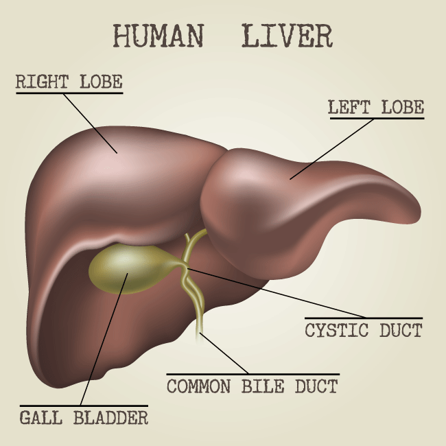
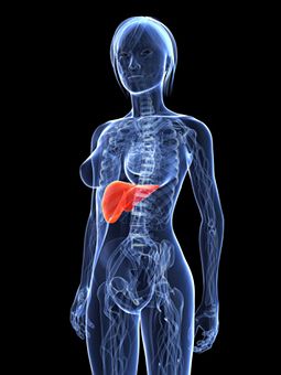
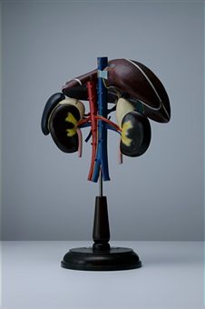
Once vitamin A has been produced, it needs to be stored until it is required. It is first converted into retinyl esters, and is then transported through the body in the lymphatic system and blood. The vitamin A that is produced from the breakdown of beta-carotene is mainly stored in the liver in the form of retinyl esters. Some retinyl esters are also stored in the kidneys, lungs and adipose fat tissue, but between 50 and 80 percent of stored vitamin A is found in the liver.

Function

Vitamin A is essential for vision. The body converts vitamin A or retinol, through an oxidization reaction, into retinal. This is combined with an opsin protein in order to produce a light sensitive molecule. When one of these molecules is hit by a photon of light, the retinal component changes its shape, setting off a sequence of events that will eventually lead to a signal being sent to the visual part of the brain through the optic nerve, where it will be decoded as vision. Retinal is responsible for the ability to detect light and therefore to see.

There are two different types of light sensitive cell in the eye, and they use different chemicals in which to see. Rhodopsin is present in the light receptors known as rods, whereas a different chemical called Iodopsin is used by the cone cells. Rods are most effective in dim light, while cones provide color vision. Both types of receptors depend upon the ability of retinal to react to light.

Vitamin A also performs some other functions in the body. It is used in the production of some glycoproteins, which are protein molecules to which carbohydrates have been attached. Vitamin A plays a crucial role in growth and bone development, reproduction and the maintenance of the skin and mucous membranes such as the lining of the mouth and nose.

These linings help to prevent infection by keeping out infectious agents from the digestive system, urinary tract and the respiratory system. The importance of vitamin A in the body is clear, since a deficiency of this vitamin can lead to abnormal development of the bones, reproductive disorders, a condition called xerophthalmia that caused the cornea of the eye to become dry, and even to death.

Deficiency

Most people will consume an adequate amount of beta-carotenes in their normal diet, but it is possible to suffer from a vitamin A deficiency when a poor diet does not provide enough vitamin A or the carotenes fro which it can be manufactured. A diet that contains low levels of beta-carotenes will not be harmful as long as enough vitamin A is being consumed in other food. A diet without enough vitamin A or beta-carotene will be harmful. If there is a deficiency of vitamin A, due to malnutrition or illness, it can be cured by eating beta-carotene rich foods.

The earliest symptoms of a vitamin A deficiency are visual problems in low light situations, dry hair and skin, fingernails that break easily and a lowered resistance to infection. The more serious signs of a vitamin A deficiency are anemia, abnormal bone development, and permanent damage to the eyes.

The retina may be injured badly enough to cause blindness. Even when there are no vitamin A deficiency symptoms, a person who is not getting enough vitamin A could suffer from an increased risk of developing diarrheal and respiratory infections and a decreased growth rate and bone development. Fertility can also be reduced.

Health Problems with Deficiency

A vitamin A deficiency can be very serious. It is rare in the US, where most people consume more than enough beta-carotene and vitamin A in their diet.

However, a deficiency in vitamin A is the most common cause of preventable blindness in children worldwide, and it affects people in more than half of the countries in the world. Low income families in Southeast Asia and Africa are most likely to be affected. It is the young children and pregnant women within these families who are most likely to suffer from vitamin A deficiencies.
beta carotene
Vitamin A is required at higher levels during pregnancy, and children have smaller stores of vitamin A in their livers than adults. An adult can have enough vitamin A in their liver to last for an entire year, but a child’s supply can only last for a few weeks at most.

During Pregnancy

During pregnancy, the highest risk of developing a vitamin A deficiency occurs during the third trimester, when both baby and mother require large amounts of vitamin A. If the mother is not receiving enough vitamin A during this period, she will suffer from night blindness, and may experience other symptoms. \

She may also have a higher risk of maternal mortality. It is not recommended for women who have a good, balanced diet to take vitamin A or beta-carotene supplements, however, since they are not necessary. Vitamin A supplements could even increase the chances of birth defects in the baby.

Deficiency in Children

Children who are not obtaining enough vitamin A are at risk of blindness and other visual impairments, and they are also more likely to catch serious diseases such as measles, or to suffer from diseases that can cause diarrhea. These illnesses can be fatal. A lack of vitamin A can also cause growth problems and defects in the development of the skeleton.

According to the World Health Organization (WHO) it is estimated that there are 250 million children of preschool age who are suffering from a vitamin A deficiency, and that between 250,000 and 500,000 of these children are made blind because of this deficiency every year. Half of the children who go blind in this way will die within the next year.

It is possible to die from a vitamin A deficiency, and according to the World Health Organization (WHO), it is possible to significantly decrease mortality rates by ensuring an adequate supply of vitamin A.

Cases in the US

In the US, cases of vitamin A deficiency are most likely to occur in patients who are elderly or who are suffering from chronic illnesses that can reduce the absorption of vitamin A and carotenes from the intestine. Patients who have inflammatory bowel disorder (IBD), pancreatic insufficiency or cystic fibrosis have an increased risk of vitamin A deficiency.

Vegans and people who suffer from alcoholism are also more likely to have a deficiency, due to decreased ingestion and absorption of vitamin A. Some cases of malnutrition do occur in the US, but these are more common overseas.

It is possible to treat a vitamin A deficiency by eating foods that contain beta-carotene or vitamin A, or by taking supplements of one of these chemicals.

If the diet contains too much beta-carotene, it can lead to the skin turning a yellowish color. It will not, however, cause an excess of vitamin A in the body. Some of the vitamin A that is produced but which is not currently needed will be stored in the liver, where it can remain for several years. Some will be stored in the fat tissue of the body. If there is a lot of beta-carotene in the diet, then it will not all be converted into vitamin A. An excess of vitamin A in the body would be harmful.

Traditional Beliefs

It is often said that carrots can help people to see in the dark. Since one of the symptoms of a vitamin A deficiency is night blindness, this belief clearly has some foundation in fact. Although eating carrots cannot enhance night vision above normal abilities it can prevent it from deteriorating by providing the precursor for vitamin A production.

Antioxidant

beta-carotene is an antioxidant. Antioxidants are chemicals that can react with free radicals, which are highly reactive, charged molecules. Free radicals are produced by the body during respiration or energy production and can cause damage that in involved in the ageing process and cancer. It may be possible for antioxidants in food to protect the body from this sort of damage, but there is no definitive scientific proof that eating foods containing high levels of antioxidants can actually help to protect the body.

Medicinal Uses

beta-carotene has been approved by the US Food and Drug Administration (FDA) as a treatment for erythropoietin protoporphyria. This is an inherited condition that is very rare. It causes problems in the metabolism of the chemical porphyrin-heme. This can lead to photosensitivity, with the skin reacting painfully to light, dysfunction of the liver, and production of gallstones. Patients are treated with an over the counter beta-carotene supplement, and may also need to take antihistamines.

Consuming beta-carotene and other carotenoids can help to prevent a vitamin A deficiency, but a diet that is rich in carotenoids is not necessary as long as there is plenty of vitamin A in the diet. There is therefore no recommended intake of carotenoids according to the Institute of Medicine’s Food and Nutrition Board.

The American Heart Association, among other health groups such as the International Agency for Research on Cancer (part of the World Health Organization) advise that people should obtain beta-carotene from a diet that is high in fruits and vegetables rather than by taking dietary supplements.

Some of the scientific research that has been conducted on beta-carotene treatment has not yet produced clear results. These treatments have not yet been proven to be successful, but neither have they been proven unsuccessful.

Potential Uses

These potential uses for beta-carotene are as treatments for cataract prevention, reducing the adverse side effects of chemotherapy, treating chronic obstructive pulmonary disease (COPD), improving cognition and memory, preventing asthma attacks that are induced by physical activity, enhancing the immune system, promoting remission in patients with oral leukoplakia, preventing and slowing the progression of osteoarthritis, treating polymorphous light eruption (PLE), reducing the risk of sunburn and UV induced erythema, and reducing complications during pregnancy. Further research may clarify the effect of beta-carotene on these conditions.

beta-carotene has also been tested as a treatment for a number of other conditions for which it was not found to be helpful.

Potential Side Effects

There was even some evidence that it could be harmful when used in patients with these conditions, particularly if beat carotene was a replacement for other more beneficial therapies. beta-carotene was not found to be effective for the prevention or treatment of Alzhemier’s disease or abdominal aortic aneurysms (AAAs), preventing the development of new moles on the skin, preventing cancer or cardiovascular disease, eradicating the bacteria that can cause stomach ulcers (Helicobacteria pylori), preventing or slowing macular degeneration, preventing stroke, or reducing injuries following surgery.

Taking supplements of beta-carotene was also found to have no effect on overall mortality rates. There is some evidence that beta-carotene may be harmful in patients who have undergone angioplasty.

Sources of Beta-Carotene

beta-carotene occurs naturally in many plants. It is found in green vegetables and fruit and vegetables that are orange or yellow. Spinach, broccoli, carrots, red peppers, nectarines, melons and mangoes are all good sources of beta-carotenes.

A diet that includes five portions of fruit and vegetables every day will provide between 6 and 8 milligrams of beta-carotene per day. A healthy body can maintain adequate levels of vitamin A with just 1800 micrograms of beta-carotene, therefore it is rare for a diet to be deficient in carotenes. There is no recommended daily allowance (RDA) for beta-carotenes, due to a lack of evidence on its importance, but the RDA for vitamin A is 0.9 milligrams per day for an adult man and 0.7 milligrams a day for an adult woman. Pregnant women and those who are breastfeeding require a higher daily intake.

Vitamin A can also be consumed directly, rather than as its precursor, beta-carotene. Egg yolks, dairy products and fish oils all contain high levels of vitamin A. Liver is also rich in this vitamin, and in fact, the liver of the polar bear contains such large amounts of vitamin A that it is poisonous to humans.

Supplements

beta-carotene is available as a dietary supplement. It can be manufactured synthetically or derived from fungi, algae or palm oil. Supplements of beta-carotene can be taken in a number of different forms, including gelatin capsules, tablets and chewable tablets.

There is some scientific evidence that consuming too much beta-carotene in the form of supplements may be harmful. A study found that there was an increased risk of developing lung cancer when people who were exposed to other risk factors for the disease, such as smoking or working in an environment where they were exposed to asbestos, took beta-carotene supplements.

There is no evidence of what the effect may be on non-smokers and people who are not already at an elevated risk of lung cancer, but it is possible that taking supplements of beta-carotene could be harmful. No similar effect has been detected when beta-carotene is eaten in food.

This means that it is important, when taking beta-carotene supplements, to avoid consuming too much beta-carotene. The UK Food Standards Agency advises against exceeding a dose of 7 milligrams of beta-carotene a day. Doctors may recommend taking a higher dose than this in some cases, but this is only when there is a serious deficiency that needs to be corrected. Anyone who is a smoker or who has been exposed to asbestos should avoid taking supplements of beta-carotene.

Absorbing Beta-Carotene

In order to absorb beta-carotene, the body must use some of the fat that has been eaten. This means that when beta-carotene is taken as a supplement, it will require some dietary fat in order to be absorbed by the body. No difference has been found in the absorption rate between individuals consuming a low fat or a high fat diet, however, since the amount of fat that is required is very low.

In order to absorb beta-carotene, the body must use some of the fat that has been eaten. This means that when beta-carotene is taken as a supplement, it will require some dietary fat in order to be absorbed by the body. No difference has been found in the absorption rate between individuals consuming a low fat or a high fat diet, however, since the amount of fat that is required is very low.

Elevated levels of vitamin A in the body can be harmful, causing problems with the bones that may lead to an increased risk of experiencing fractures in old age. Although beta-carotene is converted into vitamin A it does not pose a similar risk. The body is not efficient enough at turning beta-carotenes into vitamin A for a high intake of beta-carotenes to cause high enough levels of vitamin A to harm the body.

Food Coloring

In addition to being manufactured as a dietary supplement, beta-carotenes are produced and used as colorings in food products.

Sources:

Antioxidants

Curcumin

Curcumin is the active compound in turmeric, a popular spice often used in Indian foods and curry. Curcumin is classified as a polyphenol and it is the compound that gives turmeric its bright yellow color. Besides being a popular dietary supplement, it is used as a food coloring.

Turmeric comes from the Curcuma longa plant, a member of the same family as ginger. Turmeric is from the rhizomes of the plant, which is part of the stem that resides underground.
What is Curcumin Used For
Along with curcumin, the turmeric rhizome also contains demethoxycurcumin and bisdemethoxycurcumin. Turmeric has traditionally been used as a medicine for thousands of years in India, but more recently, it has been used to treat inflammation, cancer, and liver disease, among others.

History and Traditional Uses

The first medicinal use of turmeric comes from Ayurvedic medicine in India. Evidence suggests it has been used since at least 1900 BCE.

The rhizomes and roots of the turmeric plant, which grows wild in the Himalayas and South Asia, would be ground into a powder and used both externally and internally to treat a wide variety of ailments.

Turmeric was said to be the most beneficial for the liver, heart, lungs, and the skin.

A paste of ground turmeric was applied externally to cuts, scrapes, and burns. It was said to have a variety properties when applied externally: as an analgesic, antibacterial, antiseptic, anti-inflammatory, and astringent.

When eaten, turmeric was said to treat the following conditions:

  • Anemia
  • Epilepsy
  • Cancer
  • Diabetes
  • Food poisoning
  • Gallstones
  • Indigestion
  • Irritable bowel and other digestive disorders
  • Poor circulation
  • Staph infections
  • Irregular menstrual cycle
  • Impure breast milk
  • Low semen production
  • Fever
  • Diarrhea
  • Urinary tract infection
  • Cough and excess mucus
  • Liver toxicity

Modern Uses of Curcumin:

Today, curcumin is still used by some people for all of the traditional uses, but the long list has been expanded even further. Curcumin is used by people of all ages, but the highest demographic is people in their late 30s and older.

Curcumin is not widely used by the mass population, except some places in India, where its use originated. In North America and Europe, curcumin use is limited to those seeking natural remedies and trying to reduce modern medications in their life.

Antioxidant Properties

Curcumin has been shown to be effective as a scavenger of reactive oxygen and nitrogen in a test tube environment, but no clear results show that it works as an antioxidant inside of a living animal. Because of its low absorption rate in humans, most prepared curcumin doses are too low to even enter the bloodstream.

However, some studies have found that accumulated curcumin in the gastrointestinal tract did inhibit oxidative damage in the immediate areas of accumulation. Curcumin has also been shown to have indirect antioxidant properties by inhibiting enzymes that play a role in oxidation.

Cancer

Curcumin, in initial trials, has been shown to produce apoptosis is in a variety of cancerous cells. Apoptosis is a programmed cell death that happens suddenly without accompanying inflammation. So far, only Phase I trials have been completed in humans.

These are studies of very small groups of people. In these trials, where 3.6 g of curcumin was taken per day for 7 days, traces were found in both normal and malignant tissues of colorectal cancer patients.

In one of the few Phase II trials that have been conducted, some anticancer properties were shown in two out of the 21 study participants suffering from pancreatic cancer. Because of its low bioavailability, however, it is thought only to be effective in a liposome encapsulated form injected intravenously.

Inflammation

Although full clinical trials in animals have shown curcumin to have anti-inflammatory properties, only a very few limited trials have been conducted on humans. In one preliminary study of 18 patients suffering from rheumatoid arthritis, curcumin supplements were taken at doses of 1200 mg per day for two weeks.

Patients reported similar improvements in swelling, stiffness, and general movement that had been experienced with NSAID treatment. Another preliminary trial of 40 men recovering from hernia surgery showed that curcumin administered at 1200 mg per day for 5 days was more effective in reducing pain, tenderness, and edema than a placebo.

Two other studies showed that 1125 mg/day of curcumin, taken for 12 weeks, relieved inflammatory conditions of the eyes. Larger clinical trials are still required to make a final determination on the effects of curcumin and inflammation due to diseases like rheumatoid arthritis.

Cystic Fibrosis

Curcumin has been taken by people suffering from this genetic disease as a pain treatment. A study done on mice showed that curcumin, orally administered, can correct the abnormal protein folding and ion transportation that causes the pain in cystic fibrosis.
Treatments with Curcumin
Mice, however, which have the same genetic mutation as humans with cystic fibrosis only experience gastrointestinal effects of the disease, not the pain or lung complications associated with humans. Furthermore, a second trial based on the exact same model as the first found no benefits whatsoever.

The Cystic Fibrosis Foundation conducted one human clinical trial using curcumin, and, they too, found it to be of no benefit. The foundation, therefore, has officially recommended patients not to use curcumin for management of cystic fibrosis.

Alzheimer’s Disease

Alzheimer’s disease causes deposits of peptides to be deposited in the brain. These are called amyloid plaques. The plaques are known to cause oxidation damage and inflammation. Curcumin injections have been shown to inhibit the formation of these plaques in the body, even crossing the blood-brain barrier in animal test subjects.

Inflammation and oxidation damage seems to have been reduced. Biomarkers show that reduction may be in the areas that cause the memory deficits associated with Alzheimer’s disease. It is not certain, however, if orally-administered curcumin can cross the blood-brain barrier, which is necessary in the treatment of symptoms.

So far, clinical trials in humans have shown that 4 g/day of orally administered curcumin is safe to use. Effects on Alzheimer’s disease in humans have yet to be studied.

Curcumin Dosage and Availability

Curcumin is available today as a standardized extract, which is powdered and put into gel caps, often with other ingredients, such as black pepper or ginger. Raw curcumin is taken in doses of 3 to 10 grams, but it is usually taken as a standardized extract in doses of 300 mg to 1 gram. Doses are repeated from 1 to 5 times per day, usually with food.

Unfortunately, curcumin is very slowly absorbed by the body, so unless it is prepared with oils in food, it can take hours before it reaches the bloodstream.

The reason why curcumin is often found capped together with black pepper or piperine (black pepper extract) is because studies have shown that, when taken together, curcumin absorption is increased by 2000 percent within the first hour after consumption. In 2007 and 2008, two new forms of curcumin were manufactured: nanoparticle and lipidated. Both forms show increased performance and absorption but are still not widely available.

Studies show that curcumin itself is only found in very low concentrations in plasma and serum. Curcumin metabolites, however, are detected, but there is no evidence that the processed metabolites have the same benefits of curcumin. In fact, curcumin, taken orally, is not detected in serum at all when taking less than 4 g per day. Evidence suggests that curcumin can accumulate in the lining of the intestine where it is absorbed by the body at a later time.

Legal Status of Curcumin

Curcumin is legal as both a food additive and dietary supplement everywhere in the world. It can be found at natural food stores and, as turmeric, in open markets and supermarkets.

Side Effects

Curcumin is labeled as a generally safe product in the U.S. and no other country places a limit on its use. No serious side effects have ever been reported from high doses. Studies show that up to 12 g taken in one dose is safe, and dosage did not increase the quantity or severity of side effects. Long-term usage at 8 g/day for three months was also shown to be safe in studies.

Participants had very few, if any, side effects. The safety of curcumin in pregnant and lactating women has not been studied, but no historic evidence indicates any adverse effect. Mild side effects of curcumin experienced by some people include only nausea and diarrhea.

Curcumin Availability and Cost

Curcumin is readily available as turmeric in most food stores where it sells for about $5.00 per pound in the U.S.

As a dietary supplement, curcumin is found at natural foods and vitamin shops in a standardized extract form of usually 95% purity. Standardization claims of dietary supplements are not tested in the U.S. 100 capsules of curcumin at 500 mg can sell for anywhere between $10 and $25 per bottle, depending on the manufacturer and retail store.

Herbs

Yarrow

Where Does Yarrow Come From?

Achillea millefolium, or yarrow, is also called milfoil, sanguinary, or stanchgrass, among a long list of other names. It is capable of surviving in most soils under most situations, however, it prefers well-drained soil in direct sunlight. These plants are capable of tolerating lime, shade, and lack of water. As a matter of fact, not only do yarrow plants live longer in poor soil, but they also improve soil fertility.

Very often the health of plants growing nearby are improved and their essential oil content is enhanced, which, in turn, protects them from insects that would otherwise destroy them. Although native to Europe and Asia, yarrow became naturalized in North America as well as other countries worldwide. Throughout the eastern and central United States and Canada, yarrow is commonly found along roadsides, in old fields, pastures, and meadows.
yarrow benefits

History

In ancient folklore, yarrow, which was considered to be dedicated to the Evil One, also inherited the nicknames Devil’s Nettle, Devil’s Plaything, and Badman’s Plaything.

At one point in history, Achilles, a Greek hero of the Trojan War, used this plant to heal his soldiers, which explains why it is also referred to as Soldier’s Woundwart. It is said that he learned this remedy from his mentor, Chiron the Centaur.

After Achilles introduced this herb to the battlefield, it was used constantly up until the time of the Civil War. During this time period, the plant was crushed and applied to bullet and shrapnel wounds. Yarrow was supposed to be extremely effective in healing wounds, especially those that were caused by iron weapons. Some believed they could be protected from evil by burning yarrow on the eve of St. John’s Day.

Others believed that good health in the coming year would be guaranteed if a bundle of yarrow was tied together and hung over a doorway or over an infant’s cradle on Midsummer’s Eve. Druids used the stems to assist in foretelling the weather and the Chinese used the stems to see into the future. Many young girls burned yarrow and chanted, “Good morning, good morning, good yarrow, and thrice good morning to thee.

Tell me, before this time tomorrow, who my true love is to be,” in hopes of discovering the identity of their future husband. The importance of yarrow in ancient history can especially be seen with the discovery of a 40- to 60,000-year-old Neanderthal whose remains were found holding yarrow among other herbs.

What Is Yarrow Used For Today?

Bloodwort, another name for yarrow, is known to be a fever reducer, to have the capability to lower blood pressure, to tone blood vessels, and to be a urinary antiseptic. It is also popular for its ability to repel beetles ants, and flies. The yarrow leaves have been used to cleanse cosmetics from greasy skin and the flowers have helped to provide yellow and green dyes.

Most importantly are the compounds found in the essential oil of the yarrow plant and what healing properties they have:

  • Matricine is anti-allergic, anti-infection, soothing, and slighty narcotic.
  • Alpha-pinene is a powerful anti-inflammatory.
  • Thujone is a narcotic, but is an epileptic in high doses.
  • Achillene, Stachydrine, Marrubine is used for fevers, non-lesion bound intestinal and stomach problems,
    and is an anti-anorexic.
  • Tannins and Flavonoids are strongly anti-cancer, are HIV inhibiting, and enhance Vitamin C absorption.
  • Inuline is anti-cancer, anti-diabetic, anti-bronchitis, anti-HIV, and anti-tuberculosis. It is also a
    bronchodilator and an immune stimulator.
  • Coumarin in low doses help build blood, but it is also a narcotic and in high doses it can have an
    anti-Vitamin K effect.

Yarrow also carries along with it a broad spectrum of specific medicinal usages such as:

  • Fever -Antiseptic
  • Eye Infections -Wide spectrum antibiotic
  • Regulates menstrual cycle -Toughening footsoles
  • Gynaecological regulator -Obstetric
  • Post-birth tiredness and depression -Hemorrhoids
  • Promotes secretions of pancreas, gut, and liver -Wounds
  • Anti-diarrhea -Anti-dysentery
  • Ulcers in stomach and duodenum -Crohn’s disease
  • Involuntary loss of urine -Rheumatism, arthritis
  • Powerful spasmolytic for cramps -Nipple-care at lactation
  • Recovery of spine surgery -Paralysis without nerve section

The healing properties of yarrow don’t even stop at this list. The leaves of the yarrow plant are often smoked as a substitute for tobacco. The fresh leaves are chewed in order to relieve the pain of a toothache. In Sweden and Africa, yarrow is used to brew beer instead of hops due to the fact that yarrow is more intoxicating.

Yarrow oil is even used in most shampoos. It can also be used to open up the pores of the skin, thus helping obstructed perspiration. It is good for kidney disorders and is recommended in children when beginning symptoms of colds or the measles are present. The essence of yarrow flowers, especially the pink flowers, is good for people that need to release emotional energies that they have picked up from the environment, family, friends, and work.

Purposes That Have Been Proven Effective

It has been scientifically proven that yarrow oil is an effective anti-inflammatory, antioxidant, and antimicrobial. It is proven to be a diaphoretic, which means it increases the temperature of the body thus opening pores in order to help produce perspiration which, in turn, eliminates toxins through the skin. Yarrow can prevent or stop the growth of microorganisms, which makes it an antiseptic. It has also proven to be an antispasmodic and an astringent.

Yarrow is an emmenagogue, meaning it promotes menstrual discharge. It helps open blood vessels and is very useful in healing wounds. It has been determined to exhibit anti-tumor properties.

It is known to decrease cholesterol and to increase bile flow. It is even often used against gastrointestinal disorders and hepatobiliary disorders, or disorders which effect the liver and/or biliary tract. It has been determined to be good at healing yeast infections and boils, also. On the other hand, there is little evidence to support the fact the yarrow is an effective treatment of the common cold.

Dosages

There is no regular set of dosing standards for yarrow because it depends on how it is being used and what it is being used for. If a supplement in capsule form is being taken, most likely the dosage is to take two capsules two times a day with water at mealtimes. A handful of dried yarrow can be used to make a cup of tea in order to relieve a fever.

If equal parts of Urtica Leaf tincture, Taraxacum Root and Leaf tincture, Yarrow tincture, and Angelica tincture are combined, children can be given a maximum of ten drops (maximum dose for adults is 50 drops) up to five times a day to help relieve a fever.

For menstrual cycle regulation use 20 drops of the tincture, which can be purchased in that form, two times per day from the beginning of menses and for the life of two full cycles.

If yarrow is combined with Petroselinum Crispum and red wine, it can prevent female and male sterility. Simply used 20 drops two times dailty for two or three months. For pregnant persons hoping for a more simple labor, drink two cups of yarrow tea per day starting approximately a week before labor. To make this tea, use one handful of yarrow and two spoonfuls of Rubus idaeus. For postpartum tiredness and depression combine yarrow tincture with Cannibus sativa tincture and hops tincture. Hemorrhoids can be treated by using yarrow internally and externally.
yarrow health remedies
For internal use, take 20 drops a day. For external use, use a poultice or cream but add five grams of Calendula and one gram of Bryony tincture.

For ulcers in the stomach and duodenum, a complex regimen is needed so an herbalist is to be consulted for appropriate dosing, diet, and other herbs that may be necessary.

An herbalist also must be consulted for the treatment of Crohn’s disease and arthritis.

There is a cream made with yarrow for wounds and abrasions. For eye infections, use compresses made of yarrow, but they must be very well filtered so as to not scratch the eye.

Side Effects and Warnings

Caution should be exercised when using yarrow if the person has an allergy to ragweed. Avoid use if there are gallstones present. An allergic skin rash or skin sensitivity to light may be caused by extended use of yarrow whether it is being used medicinally or in the diet.

Using yarrow while pregnant may lead to reduced fetal weight and increased placental weight. Yarrow use may also alter estrogen activity. Women who experience heavy periods or who have pelvic inflammatory disease should not use yarrow. Even though yarrow is used to treat wounds, it still should not be used to treat large, deep, or infected wounds. The actual leaves of the yarrow plant should never be used alone internally.

Processes for Consumption

If fresh yarrow is not being used, then it must be dried. Take the whole yarrow plant, as it is all beneficial, and hang it upside down away from the sun, moisture, and dust for a few days. If the interest is for fresh yarrow, the plant flowers from May to June. Many different species of yarrow have different colored flowers, but it is best to use the white native species because the others are cultivated for their smell and color, not medicinal purposes.

The quality of yarrow can be judged simply by its smell. When making the tincture, the yarrow used can be fresh or dried. For oils and salves fresh yarrow can be used because this plant has a low water content and will not liquify the oil. Dried yarrow should be used for teas and fresh yarrow leaves should be used for poultices. The bitter taste of this plant can be covered up by cooking it into a cough syrup using something sweet such as honey.

Additional Information

Yarrow is uncontrolled in the United States. U.S. Supplement Laws regulate the sale of yarrow if it is in the form of a supplement. If it is in the form of a food or a drug, sales are regulated by the FDA, or Food and Drug Administration, and will be labeled as GRAS, or generally recognized as safe.

When shopping for yarrow, the USP, or United States Pharmacopeia, symbol guarantee safety and quality in those brands. Some popular brands of yarrow supplements include Nature’s Way, Solaray, and Nature’s Answer. Yarrow can be found throughout the internet and herbal websites as well as in local herbal stores and even some big name grocery stores that have a natural section. Prices are as low as $2.50 and simply depend on the purity and quality of what is being purchased.

Conditions

Interstitial cystitis

Interstitial cystitis (IC) is a disease that manifests as recurrent pain or discomfort in the bladder and the region of the pelvis that surrounds the bladder. The symptoms are usually different for every patient and one patient can have different symptoms with each flare up. Approximately 1 million people are affected by the condition – mostly women.

How severe the symptoms are may be different for each person as well. Some people with IC do have periods where it goes away as if in remission. Because of the differing symptoms and the fact that the symptoms are often more severe in some people than in others, it is thought that IC is actually several diseases.
internal cystitis symptoms
It may help to understand better how the bladder functions in order to fully understand what interstitial cystitis is. The bladder is a hollow organ in which the urine is stored until you are ready to empty your bladder.

When the bladder is full, signals are sent to the brain using the pelvic nerves indicating that it is time to empty. This is what produces the urge to urinate. With IC, these signals are confused and the patient may feel the need to urinate more often but smaller volumes of urine are actually emptied from the bladder.

Having interstitial cystitis can cause a number of complications:

  • It can make the walls of the bladder become stiffer, resulting in smaller capacity of the bladder.
  • Pain and the increased need to urinate can inhibit work, social activities, and other things in the patient’s daily life.
  • The constant need to urinate along with the pain can sometimes lead to emotional issues that can eventually lead to depression. Depression can worsen symptoms of any disease, including interstitial cystitis.

Alternative Names

Interstitial cystitis is sometimes called IC. It is more simply referred to as painful bladder syndrome (PBS). Scientists who research this condition also call it bladder pain syndrome (BPS). Depending on the symptoms a patient has, doctors may use the term IC/PBS. They normally do this for all cases of urinary pain that aren’t connected to any other issue like an infection of kidney stones.

Causes

There is really known definitive cause of interstitial cystitis. Much research is done and focuses mostly on the lining of the bladder (the epithelium). This layer is there to protect the wall of the bladder from toxins in the urine. According to the research, in 70 percent of patients with IC this lining contains leaks. This suggests that these leaks may be allowing substances like chemicals from beverages or food that are in the urine to enter the bladder. These substances may irritate the bladder wall, triggering the IC.

The lining of the bladder wall is made up of protective substances. Researchers who study IC also believe that some people with the condition may have lower levels of these substances. This can be another reason for the damage to the wall of the bladder found in so many cases of IC.

A substance called antiproliferative factor (APF) has also been discovered that may block the normal production of the cells that make up the lining of the bladder. Nearly all of the time that APF is found in the urine it is with people who have interstitial cystitis.

There is another theory that suggests that IC is an autoimmune disorder or that infection may be the cause of the symptoms and the bladder damage.

Symptoms

As mentioned earlier, the symptoms of interstitial cystitis may differ from patient to patient. Someone with IC may also discover that his or her symptoms change with time. The symptoms can come on or become worse with certain physical or emotional triggers like menstruation, allergies, sexual activity, or anxiety and stress.

The symptoms of IC include:

  • The continuous and pressing need to urinate.
  • Urinating frequently in small amounts. Those who have severe cases of IC may urinate as much as 60 times in a day.
  • Pelvic pain. In women the pain is between the vagina and the anus and in men it is between the scrotum and the anus.
  • Pelvic pain during intercourse.
  • Constant pain in the pelvis.

Some patients may have only the pain and others the urination symptoms. It is most common to have both the pain and the frequent urination.

Pain from interstitial cystitis can get worse if there is a urinary tract infection.

Risk Factors

There are a few factors that can put people at a higher risk of interstitial cystitis:

  • Sex – Women are diagnosed with IC considerably more often than men or children of either gender. Men can have similar symptoms that are normally associated with prostatitis.
  • Age – This disease most often occurs between the ages of 30 and 40.
  • Other chronic conditions – People who suffer from other chronic conditions like irritable bowel syndrome and fibromyalgia may be more prone to IC, however there is no known connection between these conditions.

Prevention

Any preventative measures that can be taken against a recurrence of interstitial cystitis are based on the theory that it is caused by the damage done to the bladder wall. The advice is to avoid any continuing irritation of the bladder. One way to do this is by boosting the immune system:

  1. Visit an integrative internist. These doctors focus on your overall health and the concept of preventing disease as well as treating chronic illnesses. This type of doctor will do an in-depth study of your health history. This is important since IC can occur after another serious illness like fatigue, stress, or some type of infection that may have required antibiotics.
  2. This doctor can give you suggestions for boosting your immune system. These doctors practice both traditional and alternative medicine and can give advice regarding the proper vitamins and other supplements to take for the immune system. Probiotics are often recommended.
  3. Diet is especially important. If you eat the types of food that support gastrointestinal health, this can lead to a stronger immune system. This type of diet should include plenty of proteins, vegetables, and foods that are high in fiber. Organic food is better that non-organic and processed foods.

Another way to prevent IC is to keep the pH level in the body balanced.

  1. You should have the pH in your body tested regularly. “Potential of Hydrogen” is the amount of acidity or alkalinity in any solution. By testing the body’s pH you will know if you are more alkaline or more acidic. If the pH is not balanced properly, the body is considered to be toxic. The cells in a body that is toxic are not capable of absorbing the nutrients from food or oxygen.
  2. Eat the foods that will provide a healthy pH balance. The foods you eat should be 75 percent alkaline and 25 percent acidic. Too much acidic food can make the entire body too acidic, which can cause inflammation, irritation, lower energy, weak immune systems, and interstitial cystitis.

Test and Diagnosis

Since the symptoms of interstitial cystitis are so similar to those of things like urinary tract infections, bladder cancer, endometriosis in women, and prostatitis in men, the first thing that doctors will usually do is tests to rule those things out. The diagnosis of IC is generally based on pain in the bladder combined with frequent urination and the absence of other diseases that have the same symptoms.

To rule out the other diseases, a doctor may do a urinalysis to look for the presence of bacteria; a culture of prostate secretions to determine if there is a prostate infection; or an exam to rule out bladder cancer that consists of taking a sample of the bladder and urethra for biopsy.

The doctor will also perform a complete pelvic exam and what is known as a potassium sensitivity test. In this test a solution of water and one of potassium chloride are placed into the bladder one at a time. The patient is asked to rate his or her pain and need to urinate after the placement of each solution. If the pain and urgency is significantly higher with the potassium chloride, the doctor may diagnose IC.
cystitis information

Treatment

There is no one treatment that works for interstitial cystitis in every patient. There are a number of treatments, including medications that can offer some relief but what works for one patient may not work for the next. This often results in trying several treatments before hitting on the one that is successful.

Herbal and Home Remedies

Besides following the dietary advice given in the section on Prevention, there are some other home remedies and herbal treatments that my help with interstitial cystitis.

1. Supplements and herbs

  • Calcium Citrate – Taking a calcium citrate supplement adds alkaline to urine. This can reduce the bladder irritation.
  • Gotu kola – This herb stimulates the production of the substances that make up the lining of the bladder.
  • Buchu – This is a diuretic and antiseptic for the bladder and should be taken as a tea.
  • Cornsilk – This also has diuretic properties and should be made into a tea.
  • Horsetail – This herb has properties that heal tissue as well as acting as a diuretic.

When used correctly, supplements and herbal treatments don’t usually have any side effects and can be purchased for reasonable prices at drug stores or health food stores.

2. Bladder training – This involves teaching yourself to urinate according to the time on the clock rather than when you feel the need. Start at certain intervals, like every 30 minutes whether you feel the need to or not. You can gradually up the time by 30-minute increments. This technique can help you reduce the frequency of urination.

3. Loose clothing – Don’t wear anything that put pressure on your stomach.

Pharmaceutical Remedies

There is only one prescription drug approved as a treatment for interstitial cystitis. This drug is Elmiron and it is not entirely understood how it works, but it is thought that the drug may restore the inner lining of the bladder. It can take anywhere from two to six months for you to notice a decrease in the frequency of urination. There are some minor side effects, which include slight gastrointestinal issues, and hair loss that stops when you are through taking the drug.

There are other oral medications that may help:

  1. Non-steroidal anti-inflammatory drugs like ibuprofen can relieve the pain.
  2. Antihistamines may sometimes help with the urgency to urinate.
  3. Tricyclic antidepressants can aid with relaxing the bladder to block pain.

Any minor side effects of these drugs can be found on the labels.

Surgery and Non-Surgical Options

There are some non-surgical procedures that can be done to help relieve the pain of interstitial cystitis:

  1. Electrical Nerve Stimulation – With the use of special devices either attached to the skin by wires or inserted into the vagina, electrical pulses can be sent to stimulate the nerves that go the bladder. This is known as transcutaneous electrical nerve stimulation (TENS) and is used to treat chronic pain for many conditions. It is thought that the electrical pulses enhance the blood flow to the bladder as well as make the pelvic muscles stronger that control the bladder. This treatment may also signal the body to release substances that block pain.This procedure is not very costly and takes about three to four months to produce results. Most insurance providers cover the procedure.
  2. Bladder Instillation – This procedure is also called a bladder wash and involves filling the bladder with a solution of a prescription medication called dimethyl sulfoxide. A local anesthetic is sometimes mixed with the solution. The solution is left in the bladder for about 15 minutes and then it is expelled when the patient urinates. This treatment can reduce inflammation and may even stop the muscle contractions responsible for pain as well as urgency and frequency of urination.This procedure has one side effect and it is not serious. It can cause a garlicky taste in the mouth and odor on the skin for several hours. Most insurance providers cover the procedure.

Interstitial cystitis is very rarely treated with surgery because the procedures do not necessarily relieve pain. Some of the procedures are very invasive and most of them can have complications. Surgery is usually only the option when all other treatments have failed.

The options for surgery include:

  1. Bladder augmentation – The damaged portion of the bladder is removed and replaced with a section from the colon. This makes the bladder larger. The pain usually remains. Women may often need to use a catheter to empty the bladder several times a day. The IC can also reoccur on the section of the colon that was used to make the bladder larger.
  2. Cystectomy – This is a removal of the entire bladder. There are several different methods used to reroute the urine when this is done, one of which involves attaching the ureters to a piece of the colon and cutting an opening in the abdomen so that the urine can empty into a bag outside the body.
  3. Fulguration – This procedure uses lasers to burn away ulcers in the bladder that may be causing the IC.
  4. Resection – This is similar to fulguration, but the ulcers are cut out rather than burned with a laser.

Fulguration and resection don’t always work and have been known to make symptoms worse.

A cystectomy or bladder augmentation can cost between $6,500 and $11,000. The other procedures are less invasive and cost less.

Conditions

Obesity

What is Obesity?

Obesity is a disease that is overwhelming the American population. Obesity is defined as an excess proportion of body fat to size, more particularly a weight more that twenty percent above the normal weight. Another measure to determine obesity is the body mass index, or BMI. BMI is calculated by multiplying your weight in pounds by 705 and then dividing by your height in inches twice. A healthy BMI is 18.5-24.9. One that is 25-29.9 indicates that you are overweight and a BMI of over 30 is considered to be obese.

Causes of Obesity

Generally, obesity is caused by eating more calories than you burn. In most cases, this is a result of purely eating too much while not exercising enough. While many people believe this to be the only cause, there are many other causes for obesity. The following are causes of obesity in addition to over eating:

Age

When your body ages, your metabolism slows down. This decline means that your body no longer needs as many calories to stay healthy. If you continue to eat the same amount of calories at age 30 as you did as a teenager, weight gain will be inevitable. The best way to avoid this is to lower your caloric intake and remain active throughout your life.

Gender

Females, on average, have slower metabolic rates than men do. Because of this, women tend to be more overweight than men do. In addition to this, menopause causes an additional decrease of metabolism. Men have a faster metabolism thus require far more calories than women do. Because of this, obesity in men is much rarer than in women but it does happen especially with age as the man’s metabolism begins to slow.

Genetics

Believe it or not, many studied have shown that obesity is genetic. Obesity and thinness both tend to run in families. Studies on adopted children have provided evidence for this theory. When studied, children who were adopted tended to have a closer weight to their biological parents than there adoptive parent. Even more shocking, the environment tended to not alter the trend. On average, if your biological mother is obese there is a 75 percent chance that you yourself will be obese as well. This same statistic applies to mothers who are thin as well. However, you can prevent becoming obese even if you are genetically determined to become so.

Your Environment

While genes are indeed a heavy influence on obesity, environment plays an important role as well. Environmental factors include several things. The most important of these is lifestyle behaviors such as how much you eat and your activity level. Also, busy lifestyles can lead to weight gain as well. It is much harder to prepare a healthy meal when there is no time in the day. In these situations, simply avoiding fast food can help significantly in maintaining weight.

Physical Activity

The amount of physical activity an individual receives is a major factor in obesity. Physical activities not only burn calories but also decrease appetite, especially in obese individuals. In addition to this, activities also make the body burn fat as an energy source. The majority of obesity in the world today is a result of decreases physical activity.

Psychological Factors

Psychological factors greatly influence ones eating habits and weight gain. Negative emotions such as being sad or boredom tend to cause people to eat.

Many people who are obese feel that they cannot control how much they eat and feel helpless. In these cases, an individual often partakes in binge eating in which they eat large amounts with no control.
Obese Male

Illness

There are indeed several illnesses that can lead to obesity. One of the most common of these illnesses is hypothyroidism. This is a condition in which the thyroid does not properly secrete the hormones needed.

Thyroid hormone, the main secretion of the thyroid gland, regulates the metabolism.

When this hormone is not secreted at high enough levels, the metabolic rate slows greatly putting the individual at risk for obesity. Depression can also lead to obesity because of hormone imbalances as well.

Medicine

There are certain medications that can lead to obesity. Steroids very commonly cause weight gain among individuals. In addition to steroids, antidepressants seem to have that effect as well.

Risk Factors for Obesity

There are many factors that do indeed place you at a higher risk for becoming obese. As mentioned earlier, genetics can lead to obesity. As well as heredity, gender plays an important role as well. Females are at a much higher risk for developing this disease due to the slower metabolic rate. People who work varied work shifts are also at a higher risk for developing obesity. This is because of the constant change in schedule of eating times as well as the lack of time to prepare a healthy meal. Smokers who are trying to quit are also at a high risk.

While weaning off of the nicotine, the body may crave food in order to replace the drug. You can help to prevent this by lowering your caloric intake during this process. Also, your age greatly affects your weight. People that fall in the age range of 20 to 55 are at the greatest risk for obesity. Your race or ethnic group also plays into the risk factors associated with this illness. The White population has the lowest risk followed by African Americans and Hispanics having the greatest risk.

Diagnosing Obesity

There are several measurements used to diagnose obesity. One of the most common measurements is the body mass index. As mentioned earlier, this is a measurement that tells you your proportion of body fat to your height. A BMI above 30 indicates obesity In addition to Body mass index (BMI) this measurement, waist circumference is another measurement used in diagnosis.

This measurement is taken in inches. In this test, you simply measure how many inches your waist line is. In females, the waist circumference should be less than 35 inches and males should be less than 40 inches. Any measurement above these values may indicate obesity. If either of these values are above normal, you should visit a doctor to have things further investigated.

Preventative Measures for Obesity

It is much easier to prevent the onset of obesity than it is to loose weight after words. The best preventative measure for this illness is to develop healthy eating habits as well as follow a daily exercise routine. Healthy eating habits are best developed by first being knowledgeable about healthy foods and also by keeping a food diary.

Keeping a food dairy will help you to see everything that you have eaten throughout the day and see what you need to modify. Exercise routines do not have to be a vigorous trip to the gym. Rather, participating in a daily walking routine can prove to be sufficient. The following are a few tips to help maintain a healthy lifestyle:

  1. If you are not hungry, do not eat. Many people eat when they are bored. This can pack on extra calories in a hurry.
  2. Limit the amount of fast food that you eat. If going through the drive through is un-preventable, try to order one of the healthier items such as a salad or chicken.
  3. When going out to eat, order small amounts. If you over order, you are more likely to over eat even if you are no longer hungry.
  4. Eat slowly. While eating, it takes the stomach a while to recognize that it is full. By eating slowly, you will give your stomach time to tell you that you are full before you have over eaten.
  5. Limit the amount of time that you spend on the TV, computer, or video games. During these activities you are not only sedentary but you are also likely to eat.
  6. If you are in a family situation, setting up fun activates to do together can increase activity. For example, going in the backyard and playing basketball or throwing a ball can encourage physical activity for the entire family.
  7. Keep healthy snacks such as fruits and vegetables in your cabinets at all times. While doing this, also avoid processed foods while grocery shopping. This will reduce the temptation to grab an unhealthy snack.

Treatment for Obesity

There are many different routes that you can take to treat obesity. The least evasive way is to exercise and begin a strict diet. The usual recommendation for exercise is 30 minutes a day. The generic diet prescribed is a low fat, high complex carbs (not simple), and high fiber diet. When taking this route for treatment, social support from your friends and family is key. Another route for treatment is taking medication. Because of the severe side effects of the medications used for this purpose, generally they are only prescribed when life style modifications fail to be successful.

One of the medications used is Phentermine. This particular medication is only allowed for short term use. Increased blood pressure is a side effect of this medication and it should be used with caution and with medical monitoring. Orlistat is another medication prescribed for this purpose. The medicine works by blocking the absorption of dietary fat. Unlike Phentermine, this drug can be used for a longer duration. The side effects of using this medicine include greasy stools and fat-soluble vitamins will need to be supplemented in your diet.

Surgery

As a last resort treatment, surgery can be done to help cure obesity. A gastric bypass is the most common operation done for this purpose. What this procedure does is reduce the size of the stomach and allow food to bypass part of the small intestine. By reducing the size of the stomach, you feel fuller much quicker than if it were at its original size. This alone reduces the number of calories that you intake in your meals. In addition to this, by bypassing part of the small intestines, the amount of calories absorbed is decreased as well. This method of treatment is by far the most successful. While is it indeed the most successful, this procedure should be done at an institution that specializes in this treatment.

There are many risks associated with gastric by-pass surgery and special care is needed to observe for those risks. These risks include infection of the incision, leakage from the bypass created, and pulmonary embolism. Many people also experience gallstones or some other nutritional deficiency such as anemia.

Along with the risks associated, many modifications are required in the way that you eat after surgery. Only ounces of food can be ingested at a time because of the size of the stomach. Also, food must be chewed very thoroughly or pain can result. In addition to this, you are not allowed to drink any fluids 30 minutes before you eat, during your meal, and 30 minutes after you eat. This is because there is no longer enough room to hold both solid foods and liquids. Foods such as simple sugars may need to be avoided because with the bypass, they pass though to quickly causing complications such as diarrhea and increased heart rate.

Conditions

Liver Disease

The liver is one of the most important organs in the human body. As the largest organ inside the body, it is about the size of an American football and weighs 2 to 3 pounds. The liver is located in the upper-right quadrant of the abdomen, under the rib cage. It is composed of two lobes, and each of those are made up of smaller lobules.

Oxygen is supplied to the liver from the hepatic artery, while supporting nutrients are delivered via the portal vein. The portal vein flows to the liver through the intestines and spleen, where it delivers both important nutrients for protein and cholesterol production and chemicals that need to be detoxified.

Functions of Your Liver


The liver has several functions and is considered one of the busiest “factories” in the body. Some of the main functions of the liver are to create components of the blood from raw nutrients, produce necessary enzymes and proteins, balance hormone levels, and metabolize foreign substances in the body, detoxifying it. In addition, the liver plays a secondary role in maintaining the immune system and is responsible for creating bile, which aids in digestion. Because the liver produces bile, it is not only an organ but, medically speaking, a gland. Once the bile is produced, it travels to the gall bladder for storage.
Liver Disease Treatment
There is no one ailment known as “liver disease.” Liver disease is a generalized term for several different maladies that affects the normal functioning of the liver, the loss of which can cause grievous injury to the rest of the body.

Doctors have pushed to limit the use of the term, in favor of the more specific diseases, but the term continues to persist. Liver disease is also sometimes known as hepatic disease.

The liver is a very hardy organ and the only part of the body that can regenerate itself. Even so, liver disease can cause the widespread failure of the organ once the disease has caused 3/4 of it to shut down. Before this time, the liver will still function properly.

Causes of Liver Disease

Liver disease is a broad category of other, more specific, diseases. These specific conditions that cause liver disease can work alone or in conjunction. One disease, often leads to another, more severe form.

The two most common forms of liver disease are cirrhosis and hepatitis. In cirrhosis, normal liver tissue is replaced with non-functional scar tissue that never heals. If a direct cause of the liver disease can be determined, it is possible to stop it from destroying any more of the organ. Other types of liver disease can damage the organ through inflammation, obstruction of bile, accumulation of cholesterol, decreased blood flow, foreign chemicals, or abnormal cell growth. Here are some of the diseases that are grouped into this category:

  • Alcohol hepatitis – Consumed alcohol directly affects the liver and is a poison to it, causing it to become inflamed. Fat deposits can accumulate in the liver due to chronic alcohol abuse, causing malfunction and failure. Alcohol abuse may also cause other, more severe forms of liver disease.
  • Cirrhosis – The scarring of the liver and subsequent loss of function. It is considered a late-stage form of liver disease.
  • Drug-induced liver disease – Several medications are extremely toxic to the liver and can cause permanent damage. Some of these medications can cause damage at prescribed doses, while others require an overdose or excessive use. Acetaminophen (Tylenol) is one common over-the-counter drug that can cause liver damage. Cholesterol-controlling drugs, known as statin medications, are also toxic to the liver. Other drugs that can cause liver damage are niacin, nitrofurantoin, amoxicillin, tetracycline, isoniazid, methotrexate, and disulfiram. Additionally, some natural herbs and herbal remedies can cause liver disease. Herbal or natural plants causing liver disease are ma-huang, kava-kava, and many types of poisonous mushrooms.
  • Hepatitis A – There are five types of hepatitis. Type A is a caused by a virus that is generally transmitted via accidental ingestion of feces. It causes serious inflammation that may resolve itself. A preventative vaccine exists for hepatitis A.
  • Hepatitis B – This type of hepatitis spreads through direct contact of bodily fluids (shared needles, blood transfusions, sex). It can become chronic and lead to cirrhosis or even cancer. A vaccine also exists for hepatitis B.
  • Hepatitis C – This type is also spread via direct transmission through bodily fluids. It is a chronic illness that often leads to cirrhosis or cancer. No vaccine exists for hepatitis C.
  • Hepatitis D – This is caused by a virus that is transmitted concurrently with the hepatitis B virus. The D virus cannot live without the presence of the B virus.
  • Hepatitis E – Hepatitis E can be transmitted through food and water that has been contaminated.
  • Viral liver disease – Other than the hepatitis virus, several different viral infections can cause liver damage, including mononucleosis (Epstein-Barr), adenovirus, and cytomegalovirus.
  • Non-alcoholic fatty liver disease – This disease causes fatty deposits to collect in the liver that interrupt its normal functioning.
  • Hemochromatosis – This is a disorder in which the body has a problem metabolizing iron, causing it to build up. The extra iron can get caught in the liver, among other organs, leading to inflammation and failure.
  • Liver cancer
  • Wilson’s disease – Wilson’s disease is a genetic disorder like hemochromatosis, except it affects the body’s ability to metabolize copper.
  • Gilbert’s syndrome – This is a common disease, affecting up to 7% of all people in North America. It causes abnormal metabolizing of bilirubin. Gilbert’s usually causes no major harm, no symptoms, and often requires no treatment.
  • Budd Chiari syndrome – This condition causes blood clots to build in the hepatic vein, decreasing blood flow through the organ. It can both increase blood pressure and lead to more serious forms of liver disease. Budd Chiari syndrome has several causes, including pregnancy, sickle cell anemia, and inflammatory bowel disease.
  • Congestive heart failure – While congestive heart failure is more detrimental to the circulatory system, it can also adversely affect the liver.
  • Bile obstruction – If bile becomes obstructed from the liver to the gall bladder or from the gall bladder to the intestine, the liver can become inflamed. The most common cause of bile obstruction is gallstones. Some people also have a congenital deformation in the bile ducts that can cause obstruction.

Symptoms of Liver Disease

Liver disease has several symptoms that are in common, no matter the exact cause:

  • Abdominal pain
  • Nausea
  • Vomiting
  • Jaundice – an apparent yellowing of the skin caused by elevated bilirubin.
  • Weakness
  • Fatigue
  • Weight loss

If the liver disease is caused by or develops into cirrhosis, the following symptoms can occur in addition to the general symptoms:

  • Bruising becomes easier due to a decrease in the ability of the blood to clot.
  • A buildup of bile can leave salt deposits in the skin, causing itching.
  • An imbalance of hormones may lead to gynecomastia (enlarged breasts) in men.
  • Hormonal imbalance may also cause a decrease in libido or impotence in men.
  • Rising levels of ammonia in the body may cause confusion or lethargy.
  • Decreased production of proteins caused ascites, an accumulation of fluid in the abdomen.
  • Decreased protein production can also lead to muscle degeneration.
  • Increased pressure in the liver can lead to a slowing of blood flow. This causes blood vessels to swell, leading to varicose veins along the digestive tract.

Risk Factors for Contracting Liver Disease

Several different risk factors exist that can lead to liver disease. Some of the factors can be avoided and others are genetics.

  • Liver diseases caused by direct bodily fluid contact have higher rates of incidence in people who share unsterilized needles for intravenous drug use or unprotected sex. Some people have also been known to contract hepatitis from dirty tattooing equipment.
  • Alcohol abuse is one of the leading causes of liver disease.
  • Exposure to toxic chemicals can cause a buildup of the chemicals in the liver as it tries to detoxify the body.
  • Some forms of liver disease are genetic, such as hemochromatosis and Wilson’s disease. Those with a family history of these diseases are more likely to have them themselves.
  • Acetaminophen (Tylenol) is highly toxic to the liver. Excessive use and overdosing can lead to liver disease or liver failure.
  • Taking medications that cause blood vessels to narrow can lead to liver disease. Birth control pills have been linked contracting liver disease.
  • While smoking is thought of more in its ability to cause lung-related problems, it is also attached to liver disease.

Liver Disease Prevention Tips

Some forms of liver disease are completely preventable, while nothing can be done for genetic liver disease. Here are some things that can be done to help prevent liver disease:

  • Drink only in moderation. Alcohol abuse is the leading cause of liver disease. No evidence exists linking low or moderate alcohol use to liver disease, but abuse can greatly affect the liver.
  • Minimizing direct contact with other people’s body fluids greatly reduces the chances of contracting liver disease. Any needles used to break the skin should be sterilized and not have been used by another person.
  • Always use a condom when engaging in sexual intercourse.
  • Avoid having tattoos done by anyone who is not a licensed professional. Unsterilized tattoo equipment can spread hepatitis.
  • Vaccinations for hepatitis A and hepatitis B are available and prevent transmission of the disease.
  • Fatty liver disease can be prevented by eating a healthy, balanced diet and maintaining a schedule of regular exercise.

Liver Disease Remedies

Diagnosis of Liver Disease

Since liver disease is an internal disorder, it can only be diagnosed by a health professional. Several blood tests exist to determine the presence of liver disease.

Additionally, a doctor will use a medical history, questionnaire, and observation of symptoms. Examination of the entire body is usually done as part of the diagnosis. Here are the blood tests made specifically to detect liver disease:

  • AST and ALT – These transaminase chemicals are released by the liver when it is inflamed.
  • CCT and alkaline phosphate – These chemicals are released by the lining of the bile ducts and can indicate liver disease.
  • Bilirubin – High levels of Bilirubin are released when the liver is diseased.
  • Protein and albumen – Both protein and albumen levels are affected by liver disease.

Some other blood tests are not made specifically to detect liver disease, but can be useful in the diagnosis:

  • Complete blood count – A CBC can show a low blood cell count that is indicative of late-stage liver disease.
  • INR – This detects the ability of the blood to clot, which is affected by liver disease.
  • Lipase – This can detect an inflamed pancreas that could be the result of liver disease.
  • Electrolytes, creatine, and BUN can be assessed to determine if the kidneys are functioning properly.
  • Increased ammonia levels can show liver disease.

In addition to blood tests, some physicians prefer to use imaging devices to create viewable images of the liver and other organs.

  • CT scan – This is a computerized axial tomography image of the internal organs.
  • MRI scan – An MRI is a magnetic resonance imaging technique.
  • Ultrasound – Ultrasound uses sound waves to create an image of internal organs. This is commonly used on the bile ducts and gallbladder.

The last procedure that is used in determining liver disease is a liver biopsy. A long needle is inserted through the chest to pull out a small sample of liver tissue for examination. This can be used to help determine a specific liver disease once general liver disease has been diagnosed.

Treatment of Liver Disease

Since liver disease has many different causes and is made up by many different specific diseases, treatments can vary greatly. Hepatitis A treatment consists of keeping the body hydrated while the immune system does most of the work in clearing it up. If gallstones are the cause of the liver disease, passing them will relieve the disease and the liver will heal itself. If they cannot be passed, then surgical removal is necessary.

Patients with more severe forms of liver disease, such as late-stage cirrhosis will need medications to control the disease and maintain function. Protein absorption is big problem and part of the treatment for cirrhosis. To minimize water retention, a low-sodium diet is prescribed. If fluid does accumulate, it may have to be removed through paracentesis, which requires a needle be inserted through the abdomen and the fluid pulled into a syringe.

Surgery may be required for people with liver disease in cases where portal hypertension is present. This type of blood pressure increase can be relieved through one of two surgical operations:

  1. Transjugular intrahepatic portosystemic shunt (TIPS) – This surgery requires a stent be placed in the center of the liver to connect the hepatic vein with the portal vein.
  2. Distal splenorenal shunt (DSRS) – This surgery relieves pressure by connecting the part of the vein that goes to the kidney with part that runs from the spleen.

In the final stages of liver disease, when failure is imminent, a liver transplant may be necessary. Before a transplant can be performed a thorough medical work-up must take place to determine if the transplant is possible and from whom a liver can be used. Many people require a liver transplant, but livers are only viable for a short time after death. In some situations, a living donor can be used in a procedure that removes up to one-third of their liver and puts it in the patient. There are currently more people that require new livers than new livers available. This means patients are placed on a waiting list. In some cases, when no liver is available for a length of time, the patient may die before he or she reaches the top of the list.

If a liver transplant becomes available, the procedure takes anywhere from six to twelve hours to complete. Even if the operation is successful, the patient’s immune system may still reject the transplant as a foreign body. Some medications may help the body assimilate the transplanted liver, but there are no guarantees. If everything goes well, a liver transplant patient will be allowed to go home after two or three weeks of recovery.

Home Remedies for Liver Disease

Although liver disease is a serious ailment that should first be cared for by a medical professional, several home remedies exist that can help to alleviate liver disease by relieving symptoms and cleaning impurities from the liver. The most important home remedy is to eat a balanced diet. A diet rich in lean protein, such as provided by eggs and fish is extremely important. Most fat should also be removed from the diet. In addition, fresh vegetables are important, especially carrots, spinach, papaya, and cucumber. Other vegetables important to healthy liver function are cabbage, mushrooms, sprouts, tomatoes, beets, and beans. These fresh vegetables can help to detoxify the liver, relieving symptoms. Here are some other home remedies for liver disease:

  • Avoid alcohol and caffeinated beverages.
  • Yoga and breathing exercises can help to get proper amounts of oxygen to the liver. Some forms of yoga also provide a massage to the liver that is beneficial in relaxing it and keeping it working smoothly.
  • Drink plenty of fresh, clean water.
  • Avoid salt and refined sugar.
  • Drink one glass of fresh spinach/carrot juice (1:1 ratio) per day.
  • Dandelion root tea has been shown to be beneficial to the liver.
  • Licorice root can provide some benefits to those with liver disease.
  • Milk thistle, turmeric, and guduchi are herbs that have traditionally been used to keep the liver clean and functioning properly.
Conditions

Jaundice

Jaundice is a yellowish discoloration of the pigment in the skin that results in several other problems as well. The condition is most commonly found in newborns and infants but can affect adults as well. The term “Jaundice” comes from the French language and means “yellow”.

Alternative Names For Jaundice

The most common alternative name for jaundice is Icterus. Other terms are Yellow Skin and Yellow Eyes. There are also several different types of jaundice, each of which is given a distinct name. Neonatal Jaundice is the most common form and occurs in newborns a few days following birth. Hemolytic Jaundice is caused by the body’s red blood cells being destroyed or broken down. Hepatocellular Jaundice is the result of a dysfunctional liver, or a liver that is unable to properly produce bilirubin.
Jaundice Types
These are the most common types of jaundice but other rare forms may exist and have only been rarely documented.

Symptoms That May Accompany Jaundice

The most common symptoms of jaundice are a slight yellowing of the skin and a similar yellowing in the eyes. For most infants, parents will first notice the discoloration in their baby’s face. The yellowing will continue downward beginning in the chest and then falling towards the arms, stomach, and legs. Infants may also experience some of the following symptoms as well:

  1. High-pitched or drawn out screams.
  2. The infant may always seem sick, sleepy, and difficult to wake up.
  3. Slow-weight gain.
  4. Other signs such as irregular bowel movements and a high fever.

Adults will have similar symptoms as an infant when dealing with jaundice, but may also notice a few other signs. Jaundice will often be accompanied by the urine becoming a brown-yellowish color. Stools may become more pale or white in adults who have jaundice. Because jaundice denotes to there being a problem in the liver and digestive system, the person may also experience vomiting, diarrhea, nausea, and a loss of appetite.

Causes Of Jaundice

Jaundice is typically the result of too much bilirubin being produced so that the liver cannot remove it from the bloodstream. Hemoglobin is a chemical which is found within all red blood cells and is beneficial to the survival of the cells. When red blood cells become old or not useful, the body will destroy them and release the iron that is found within the Hemoglobin. Bilirubin is produced as the product of the iron being released from the Hemoglobin. If too many red blood cells are dying and too much bilirubin is being produced, the body may not be fully capable of dealing with so much bilirubin.

The production of too much bilirubin may not always be the culprit. Often times the liver will have a small defect that prevents the bilirubin from being removed from the blood. Other times a blockage may arise within the digestive system that blocks the flow of bile and bilirubin tot he intestines. This will cause less flow of the bile and can result in a form of jaundice that is related to a disease called cholestasis.

In the case of infants, jaundice is caused by a slightly different problem. A newborn infant will have a very high amount of red blood cells whose life cycles are relatively short. Because of this high turnover rate of red blood cells, infants may have excess bilirubin during the first few weeks of life. Other causes of jaundice in newborns can include:

  1. Some type of internal bleeding or hemorrhage.
  2. A blood infection.
  3. Some viral and bacterial infections can cause the skin to turn yellow, similar to jaundice.
  4. A dysfunctional or malfunctioning liver
  5. Some form of abnormality in the infant’s red blood cells.

The term jaundice refers to the actual “yellowing” of the skin and eyes, but is not a disorder in itself. Jaundice is typically a sign that there may be a problem in the liver or digestive system.

Risk Factors That Can Be Related To Jaundice

Premature infants are at the greatest risk of having jaundice. Babies born before 37 weeks are among those with the highest risk of having jaundice. Infants born after 37 weeks may still have jaundice but are at less of a risk. The reason that prematurely born infants have a greater risk of jaundice is due to the liver being less developed.

Babies who are born with a blood-group incompatibility are also at risk. Incompatibilities issues arise if your baby has a different blood type than your own. Babies who receive intense bruising during birth may have a higher level of bilirubin in their body due to the destruction of more red blood cells. Thus, bruised babies may have a greater chance of having jaundice. Children who are breast-fed have a higher risk of jaundice, especially if they are having a difficult time nursing and are not receiving the proper amounts of nutrition.
In the case of adults, those who have had some form of Hepatitis are generally at the greatest risk. Also, people who have alcoholic liver diseases or are heavy drinkers are at a risk of jaundice. Persons infected with the HIV virus or other sexually transmitted diseases may be more susceptible to having jaundice. The recipient or donor of a recent liver transplant or other operation on the liver may also show signs of jaundice.

Prevention Tips To Help Avoid Jaundice

Because jaundice occurs in over 75% of newborns, it can be difficult to prevent. While in the womb, the baby relies on the mother’s liver to perform most of the reduction in excess bilirubin. When the baby is born his or her liver may not yet have had the practice to do this on their own. With time their own livers will be able to remove excess bilirubin.

The best way to prevent jaundice from becoming more sever is to ensure that your infant is getting the nutrients he or she needs by properly breast feeding them. Infants should be fed 8 to 12 times a day on average and be fed approximately every two hours.

Children should be examined within 5 days of birth by a trained children’s physician. The child’s doctor may be able to note signs that suggest jaundice before the parents can and thus prevent the jaundice from becoming worse. Also, make sure to check your infant’s urine and stool. Urine should be odorless and clear, if it is a yellowish color then that may be a sign of jaundice. A stool that is pale, pink, or purple may be accompanied by jaundice. Healthy stools range in color from green to yellow.

Adults can prevent jaundice by avoiding those activities which may be related to some of the above mentioned risk factors. Adults who drink excessive alcohol and contract liver disease may have jaundice. One of the best ways to prevent jaundice in adults is to avoid alcohol. Adults should also avoid any sexual activity, such as unprotected sex, that may put them at risk of a sexual disease. Do not allow yourself to be poked by an unclean needle. If you have recently had an operation that has affected your liver it is important to follow all of the surgeon’s advice in order to care for your liver and prevent jaundice.
Jaundice Testing

Tests And Other Diagnosis Considerations When Dealing With Jaundice

Infants will commonly receive blood and urine tests that can determine whether or not they have jaundice. The most common test is called the “Bilirubin Test”. The Bilirubin Test is simply a test of the blood to determine if bilirubin levels are too high.

Adults who take this test should not eat or drink for 4 hours prior to appointment. Levels of bilirubin in the blood is typically between .20 mg/dL to 1.5 mg/dL, anything that is significantly higher than these amounts should require a physician’s attention

Adults who have signs of jaundice that may denote a more severe health problem may be more fully examined with the aid of ultrasound techniques, CT and MRI scans, or an endoscopic retrograde cholangiopancreatography. These scans are used to examine organs, such as the liver, and determine any malfunction.

The scans can also be used to uncover any blockages near the liver that are preventing excess bilirubin from exiting the body. A liver biopsy, in which a small piece of liver tissue is extracted and examined, may also be used in order to determine if the liver is not performing its proper function.

Treatment Options

Herbal And Home Remedies Commonly Used To Treat Jaundice

Several herbs have been used for hundreds of years to help buildup a strong liver. These herbs can also help fight against jaundice, but should be used accompanied by medical care and not by themselves. Some commonly used herbs include:

  1. Bitter Luffa
  2. Radish Leaves
  3. Tomatoes
  4. Snake Gourd Leaves
  5. Pigeon Pea Leaves
  6. Sugarcane Juice
  7. Lemon Juice
  8. Barley Water
  9. Jaundice Berry (also known as Berberis vultaris)

Most of these herbs can be found at a local Herbologist’s or Naturalist’s office, and some can be found at general nutrition centers and department stores.

Proper home care of jaundice is best provided by receiving adequate physical rest. Recovery is dependent upon the liver being able to handle the amount of bilirubin from destroyed blood cells. Blood cells are destroyed less when a person is sleeping or resting than when they are physically active. Patients should drink plenty of juice of all kinds throughout the day and be on a light carbohydrate diet..

Pharmaceutical Treatments Of Jaundice

Infants who have severe jaundice will often be recommended to use phototherapy. Phototherapy is the use of light waves to eliminate excess bilirubin in your baby’s blood. The skin and blood will absorb certain light waves and can use the energy of the light to change the bilirubin into more useful products that their body can either use or pass through their digestive system. Phototherapy is relatively inexpensive and special “biliblankets” can be used at home in order to avoid excess hospital bills. Phototherapy can often cause frequent or loose bowel movements, but this is normal. If these bowel movements persist for an extended period of time following the treatment of jaundice you should contact a physician.

Because jaundice in adults is usually related to some other disorder or disease, physicians will typically not prescribe antibiotics or other medications unless they are used to cure the related disorder.

Surgical Treatments For Jaundice

Gall stones can often be the cause of jaundice in adults. In this case, a surgeon may opt to remove the gallstone and remove any excess bile from the patient. Blood transfusions can be performed on infants and adults who experience sever jaundice. Aside from these cases, surgery is not necessary in the treatment of jaundice. In the case of a sever liver problem that is also accompanied by jaundice, the patient may be required to have a liver transplant.

Conditions

Corns

Corns are hardened areas of skin that are the result of constant rubbing and pressure placed on specific areas of the body, most commonly the feet. These areas can become painful and bothersome for some people.

Symptoms

Corns and calluses are skin that becomes thick and hardened. These areas of skin may also appear flaky and dry at times. These hardened, thick skin areas are most often found on a person’s hands, feet and other areas of the body that may suffer from being consistently rubbed or pressed.

Causes

Typically corns and calluses are caused when a person subjects certain areas of their skin to pressure or friction from consistent use. Corns are usually skin that thickens on the top or side of a toe. A common cause for this happening is shoes that do not fit the person’s foot properly. Calluses are areas of thickened skin that develop on the hands or the soles of a person’s feet. This typically occurs from performing physical labor tasks.
Remedies for Corns
The thickening of the skin occurs as a protective reaction to help prevent the development of blisters that can be painful and troublesome. People who have bunions on their feet most times will develop a callus over the bunion due to the bunion sticking out and rubbing against the shoe.

Risk Factors

Whenever a person consistently subjects parts of their body to repetitive rubbing or pressure, the possibility of developing corns or calluses is present. There are certain things that do put people at a higher risk factor for developing a corn or calluses. This includes:

  • Age – As a person ages, their feet tend to become wider and flatter. The soles of their feet also tend to lose fat on the padding of their soles. This the feet to fit improperly in the shoe and allows for rubbing to occur. These problems also lead to improper pressure being put on areas of the feet that will cause skin to harden and thicken up.
  • Cosmetic Foot Surgery – When a person has cosmetic surgery performed on their feet for situations such as to be able to fit into dress shoes easier, or to reduce the size of a person’s foot, this can lead to foot problems. Since the foot is designed to support a person on various pressure areas of the foot, reducing the size of a foot can throw the body’s sense of balance points off. This will lead to the foot having added pressure points in areas that it is not use to leading to the development of calluses and corns.
  • Gender – Studies that have been conducted show that women are at a higher risk of developing corns on their feet. This is normally due to the types of shoes women tend to wear. This is also a result of women wearing a variety of different types of shoes for dress, exercise and causal wear without allowing their feet to adjust to just one or two different pairs of shoes.
  • Occupational Risk Factors – The type of job and the environment in which a person works has a lot to do with their risk of developing corns. A person that is on their feet a lot or who perform a great deal of physical labor are at a greater risk of developing corns and calluses than someone who sit’s at a desk for the majority of their work shift. One example of this is a farmer who spends their time tending to their crops. Almost one hundred percent of their job requires physical labor. They develop corns and calluses to help prevent painful blisters from occurring.
  • Pregnancy – Pregnant women are at a higher risk of developing corns and calluses because of the additional pressure placed on their feet that is not normal. The feet have specific points that it is used to receiving the majority of the pressure through regular wear and this is thrown off during a pregnancy.
  • Sports and Activities – Since the majority of sports and extracurricular activities, such as dancing, require a person to spend a great deal of time on their feet while being active. These activities increase the likelihood of your feet and other parts of the body to experience extensive pressure and rubbing, causing corns and calluses to develop.
  • Weigh Gain and Obesity – People who have gained weight or who suffer from obesity are at an increased risk of developing corns and calluses because of the extra pressure that they are putting their feet under to support the extra weight.

Prevention Tips

Corns can be very painful. There are cures to help remove corns that have developed. However, there is no guarantee that corns will not return or develop in another area. The best thing to do is prevent them from occurring in the first place. Here are some tips and techniques to help reduce the chances of developing corns.

Wear socks that are clean and dry. Providing continued moisture to your skin can erode the skin’s natural toughness. This is when corns will develop. Socks lock in moisture to the foot without allowing them to breathe. Wear socks continuously also contributes to foot odor and conditions such as athlete’s feet.

Shoes should not be worn indoors. Unless you are wearing a special corrective foot wear for a orthopedic problem, you should try to go bare foot around your home as much as possible. This gives your feet a break from the constant pressure and rubbing that occurs when wearing shoes.

Shoes With a Natural Curve

Shoes that come to a point, are thin or straight in design or that have a flat foot appearance, are more likely to cause rubbing and unnatural pressure points to your feet when wearing them. This can lead to the development of corns.
Do not wear shoes that are tight in the toes. Shoes that have a tight fit in toe area will definitely rub against the inside of the toe the entire time they are worn. This will result in a corn developing on the toes. Opt for wider toe shoes that allow for comfort and are natural fitting.

Check inside seams of shoes. If shoes have inside seams, make sure that they will not rub against the toe while wearing. Shoes that do not have protruding seams are less likely to contribute to developing corns. The more padding the better. When purchasing shoes, look for styles that have extra padding to help create a buffer for your feet and toes. When your foot settles into this buffer, it will maintain its natural position and result in less rubbing and pressure. You will also want to pay special attention to the ball of the foot and the back of the heel area. This is a common place for corns to develop.

Podiatrist Recommendations

Talk to a podiatrist about inserts. If you are unable to find shoes that fit your feet properly, talk to a podiatrist about getting inserts for your shoes. There are special pads, shoe inserts and wedges that can be specially made to your foot’s natural design to help eliminate the chances of developing corns as well as other painful foot conditions.

Surgery may be necessary. If you are unable to deal with the pain of an existing corn or you have a severe deformity of the foot, you may need to speak with a podiatrist about surgical treatments that are available that can help you deal with painful corns. This should always be a last resort when dealing with corns.

Test and Diagnosis Considerations

When diagnosing a corn, doctors and medical professionals will perform a visual inspection of the area and provide you with treatment options. They may inform you of some medicines or supplies available to treat corns. Doctors may even discuss surgical options that you have to remove the corn.

Treatment Options

There are many treatment options available to help patients deal with corns. There are medicines and over the counter medical supplies, surgical options that can be performed by a surgeon and countless natural remedies and cures available to try. Here are some of the options that a person has to help deal with painful corns.

Medicines and Over The Counter Medical Supplies

Special pads that are designed to place over the corn can be purchased over the counter. These pads can help to prevent future rubbing and pressure to the corn. Some varieties of pads available are medicated, meaning that they have an acid medication designed into the pad itself to help dissolve the hard tissue of the corn. This does help, but the medicine itself can harm good skin as well as the corn if it is not positioned correctly on the corn.

Shoe padding and shoe inserts are available. These products are meant to act as a bunting for your foot, allowing it to cradle around your foot and mold it into place to prevent rubbing and pressure points.

Over the counter acid medications are available. This usually comes in a liquid or gel form and is meant to be applied to the corn to soften the toughened skin. This should not be used by people who have problems that do not allow them good feeling in their feet areas. This is also not a recommended option for a person with pre-existing health problems like diabetes.

There are specially designed tools available to help shave away the tough skin of a corn. These tools are not recommended for people who have a decreased ability to fight off infections or who do not heal well, such as diabetics.

Pumice Stones

Pumice stones are a great over the counter option when trying to eliminate a corn. Soak your feet for approximately 20 minutes. Rub the pumice stone over the corn until you have removed as much dead and dried skin as possible. Continue this method for as many days as needed without making the area around the corn raw. Diabetics should not use pumice stones because of the chances of breaking the skin and causing infection.

Surgical Options

Surgeons can cut off the corn or callused area to help eliminate the pain. If the corn is caused by a toe that is not straight, surgeons can straighten the crooked bone to help reduce the likelihood that it will return. If the corn is the result of a toe that is too long or too short and causes rubbing, surgeons can help to shorten the bone or lengthen the toe using cosmetic surgery on the feet.

Doctors can burn the corn off using a medical acid treatment, similar to the treatment used for the removal of warts.

Natural Cures and Remedies

Fill a basin or tub that can be used to soak your feet in, with as hot of water as you can stand without getting scalded. Pour in about half a cup of Epsom salt into the water filled basin or tub. Soak your feet for 20 to 30 minutes twice a day. Continue this treatment for approximately one week. During the time of treatment, avoid wearing socks and shoes as much as possible.

Dip a cotton swab into castor oil and apply to the corn. Using medical adhesive tape, tape the cotton swab to the corn and allow it set over night. Continue this process for one week and you should begin to notice a big difference in your corn.

Some patients claim that if you tie a slice of lemon over the corn and leave it there over night you will get results.
Grind three or four licorice sticks and mix with half a teaspoon of sesame oil or mustard oil until a paste like substance forms. Apply this paste to the corn and leave on overnight. This will gradually soften the skin and reduce the size of the corn.

Grind chalk into a powder and mix with a small amount of water to form a paste. Apply the paste to the hardened corn and leave on overnight. This will soften the corn and shrink the size of it.

Conditions

Appendicitis

Appendicitis is an inflammation of the appendix. It causes severe pain in the abdomen and can be treated surgically. An appendectomy is an operation to remove the appendix.

The appendix is a small tube, shaped rather like a finger. The average appendix is approximately 4 inches long and about 0.31 inches in diameter. It is closed at one end, and at the other is attached to the cecum, a pouch shaped part of the colon of the large intestine. This section of the large intestine is near to the place where it is joined to the small intestine. Food travels from the stomach, through the small intestine and into the large intestine, passing the appendix on its way.

Type of Organ

The appendix itself is usually considered to be a vestigial organ, that is one that no longer has any function. Some research points to possible secondary functions for the appendix, but it is not an essential organ, and it is possible for a human to live a perfectly healthy life without an appendix. One secondary function that has been suggested for the appendix is that it could provide an environment in which useful bacteria can grow, and perhaps one in which they can survive when illness, specifically diarrhea, flushes bacteria out of the digestive system. Another suggestion is that the appendix could play a role in the immune system, since it usually contains many lymphoid cells, which are involved in the fight against infections.

Appendicitis is a common condition that affects one in every fifteen Americans. It usually presents in patients between the ages of 10 and 30, and is very rare in children under 2 years of age.

Medical Emergency

Appendicitis is a medical emergency that requires immediate attention. If left untreated it can be fatal, since the inflammation can increase until the appendix bursts, releasing the contents of the intestine and infectious material into the abdomen. The resulting infection, known as peritonitis, requires strong antibiotic treatment in order to save the patient. If left untreated, the patient can go into shock and die.

Abscesses, which are pockets filled with pus, can sometimes form on the outside of an infected appendix. These must be treated before the appendix can be removed. If the abscess tears, the pus will leak into the abdomen, where it could spread the infection.

In some cases, the infected appendix can be sealed off from the abdomen by scar tissue produced by this type of abscess on the outside of the appendix. This can prevent peritonitis, since it becomes impossible for infectious material to be leaked from the appendix. It is not possible to recognize an abscessed appendix before conducting surgery, however, therefore all cases of appendicitis must be treated as emergencies with the potential to develop into peritonitis.
Appendicitis Symptoms

Symptoms

The first symptom of appendicitis is a dull pain that occurs near the upper abdomen or navel. It then moves down towards the lower right of the abdomen, becoming a sharp pain over a period of several hours. The pain generally increases over approximately 12 to 18 hours, becoming very severe. The patient will often be unable to find a comfortable position or to sit still, due to the pain. In some cases, the pain may occur in a different part of the abdomen than would usually be expected, particularly when the patient is a child or a woman who is pregnant.

Other common symptoms are:

  • Nausea or vomiting, soon after the abdominal pain begins
  • Loss of appetite
  • Swelling of the abdomen
  • A fever (99 to 102 degrees Fahrenheit)
  • An inability to pass gas

Some patients may also experience other symptoms. In approximately half of the people who have appendicitis, there will also be a sharp or dull pain elsewhere in the abdomen, the back or the rectum, cramps, diarrhea with gas, constipation, pain when urinating, or vomiting before the abdominal pain begins.

Anyone who is experiencing the symptoms of appendicitis should seek medical attention as quickly as possible. Many people begin by making an appointment with their family doctor, since their symptoms have not yet become severe. If the doctor suspects appendicitis, they will then be referred on to a surgeon. If the pain has become severe, then the patient is likely to seek help at the emergency room instead, and be transferred to surgery from there.

Diagnosis

If a patient arrives at the hospital or a family doctor’s offices with abdominal pain that may be appendicitis, the doctor will want to know how long the pain has been there, how severe it is, where exactly in the abdomen it is, and whether there has been any change in the pain since it first began. They will also ask whether there have been any other symptoms.

A doctor will perform a number of diagnostic tests when a patient presents with some or all of the symptoms of appendicitis. A number of other conditions can cause similar symptoms, therefore it can be difficult to correctly make the diagnosis, and the patient may not be able to recognize the condition for themselves.

The doctor will physically examine the abdomen to check for any inflammation. They will probably press down upon the abdomen and ask the patient whether there is any change in the pain. In cases of appendicitis, the pain increases when the pressure is released. This happens because the peritoneum is inflamed. The doctor will also check for any rigidity in the abdomen, and whether the patient’s muscles are reacting to the pressure by becoming tense. This is known as guarding, and is common during appendicitis.

They will usually also perform a rectal exam and conduct tests on the blood and urine to check whether the body is fighting off an infection and to rule out the possibility that the patient is actually suffering from a urinary tract infection. A high count for white blood cells in the blood test implies that the patient has some sort of infection. Urine tests can identify urinary tract infections, and if there is are red blood cells in the urine when it is examined under a microscope, it suggests that the pain is being caused by kidney stones rather than appendicitis.

A CT scan or an ultrasound may also be used to investigate the abdomen. This can confirm the diagnosis of appendicitis, or find any other reasons why the person could be suffering from abdominal pain.

It is usual for the doctor to recommend surgery whenever appendicitis is suspected, even if they are not entirely certain that the patient is suffering from this condition, since it is vital to operate as quickly as possible if the condition is present.

Causes

Appendicitis occurs when the appendix is blocked. This blockage can be caused by stool, a foreign body, cancer, or swelling of the appendix due to an infection.

Risk Factors

Anything that increases the chances of a blockage that could affect the appendix can increase the risks of developing appendicitis.

There is no way to prevent appendicitis, but when the symptoms do occur, it may be possible to reduce the chances of the appendix rupturing. The best way of preventing this is to get medical attention before the appendix can burst, but there are certain things that anyone with suspected appendicitis should avoid, since they can cause an inflamed appendix to rupture. They should not eat or drink anything, take any pain medication, laxatives or antacids, or use a heating pad.

People who eat plenty of fiber are less likely to suffer from appendicitis, so a diet rich in fruits and vegetables may reduce the chances of developing this condition.

Treatments

Appendicitis is treated by surgical removal of the appendix. A dose of antibiotics is usually given before the operation in order to protect the patient from the possibility of peritonitis developing.

If the appendix has abscessed, then the patient may need to have an operation to drain the pus before the appendectomy is performed. The pus is drained through a tube that is inserted into the abscess. The patient will usually need a few weeks to recover from this procedure before the appendix can be removed. Patients who have an abscessed appendix will therefore require two separate operations.

If the appendix has burst, then treatment with antibiotics will also be necessary in order to cure the resulting infection, which is known as peritonitis, since the lining of the abdominal cavity is called the peritoneum.

There are two ways in which an appendectomy can be performed. Both require a general anesthetic, which will put the patient to sleep while the surgery is performed.
Appendicitis Treatment
An open appendectomy requires an incision of about four inches in length to be made in the abdomen, through which the appendix is removed. If the appendix has ruptured, or if there is an abscess on the appendix, then an open operation will probably be necessary, since it provides the surgeon with the opportunity to clean out the abdominal cavity.

A laparoscopic appendectomy is conducted through a number of much smaller incisions in the appendix. This technique uses a laparoscope, which is a thin instrument with a camera on the end of it that can be inserted into the abdomen and used to find the appendix and provide a view inside while performing the procedure. Since the incision is smaller, patients whose appendectomy was laparoscopic will usually recover from the operation faster from those whose operation was open.

After the surgery, the patient will usually need to take pain medication while the incision is healing. There are no alternative therapies that can help to treat appendicitis, but there may be strategies that can help the patient to cope with the pain they experience while recovering. Some patients benefit from techniques such as guided imagery while dealing with postoperative pain. This involves imagining a favorite place in order to create a distraction from the pain. Other activities such as listening to music, watching TV, reading or talking with a friend, can also help patients to forget about the pain.

There is some recent evidence that antibiotics alone may be an effective treatment in some cases of appendicitis, without the patient having to undergo surgery. This treatment is currently being tested in clinical trials. It depends upon recognizing which patients are at low risk of the appendix rupturing, which there is not currently a reliable means of doing.

Traditional Remedies

There are no traditional cures that can treat appendicitis, but there are some remedies that have been used in the past to cope with this condition. None has been scientifically proven to be effective, and it is essential that anyone who suspects they have appendicitis visits their doctor and receives proper treatment.

In Traditional Chinese Medicine, certain herbs are believed to be able to treat appendicitis. A scientific study was conducted on 425 patients with appendicitis. They were given herbal preparations from Chinese medicine. Some patients also received antibiotics. Most of the patients who were included in the study experienced an improvement in their condition, without having an appendectomy, but 30 of the patients relapsed with appendicitis soon afterwards. Although these results are interesting, and may suggest possible research topics for the future, surgery is still the only option for curing appendicitis.

In addition to herbal remedies, acupuncture and electro acupuncture have been used in Chinese medicine in order to treat appendicitis. No clinical trials have been conducted to test the efficacy of these treatments.

Outside of the Chinese tradition, other cultures have attempted to treat appendicitis with herbal remedies. Homeopathic remedies for the condition include Bryonia and Belladonna. Indian ayurvedic medicine suggests a number of remedies for mild cases of appendicitis, including drinking buttermilk, carrot or beetroot juice, or water in which fenugreek seeds have been boiled. There is no scientific evidence that any of these herbal remedies work for appendicitis.

Traditional remedies are unlikely to help in cases of appendicitis, although some may help to improve the digestion or to alleviate some of the symptoms. In ayurveda, for example, ginger is suggested as a remedy for nausea and vomiting.

Alternative therapies for appendicitis probably only exist because of the difficulty in diagnosing the condition, which may have led to supposed cures when the patient did not in fact have appendicitis. Practitioners may also have believed that they cured appendicitis when the condition was very mild and resolved itself, with no help from their remedies. This could have led to the treatment being passed down over the generations as a remedy that was believed to help people with appendicitis.

Prognosis

If the condition is treated quickly, with an appendectomy and antibiotic treatment if the appendix has burst, then the prognosis is good. The patient should expect to spend a day, or possible two days, in the hospital after receiving the surgery. It will take up to three weeks for the patient to recover from the operation, but there should be no further problems and they will be able to lead a completely normal life, without their appendix. Open surgery requires a longer healing period than laparoscopic surgery. The patient will also take longer to recover from their appendicitis if the appendix burst before it could be removed.

Patients who have had an appendectomy should contact their doctor if they experience any dizziness or faintness, uncontrolled vomiting, blood in their urine or vomit, increased pain in the abdomen or the incision, redness or pus in the incision.

During the recovery period, the patient can take a number of measures to help themselves to heal and to avoid complications resulting from the surgery. They should avoid any strenuous physical activity. If the procedure was performed laparoscopically, they will need to limit their activity for between three and five days after the surgery. If their operation was open, then they should avoid any physical strain for ten to fourteen days afterwards. The recovery time will vary between patients, but the doctor will be able to advise on when each patient can return to their normal activities.

While the patient is recovering, they should be careful when they are coughing, laughing or otherwise placing a strain on their abdomen. They may feel some pain when doing any of these things. The abdomen can be protected during these actions by placing a pillow across the abdomen and placing pressure upon it in order to brace the stomach.

Once the patient is ready to begin moving around again, they should begin gradually and start with short, sedate walks. It is important that the patient does not try to push themselves too hard before they are ready to return to their normal lifestyle. The doctor will be able to advise the patient on when they should expect to go back to work or take up other activities again. Children who have undergone an appendectomy are usually able to return to school after about a week, as long as they avoid any particularly strenuous activities such as sports and gym classes, which they should usually avoid for between two and four weeks after surgery.

While they are recovering, the patient should ensure that they get plenty of rest and sleep. The healing process can use up a lot of energy, and they may require more sleep than they normally get, and could feel sleepier than usual.

The patient will probably be taking some pain medication during the recovery period. If this is not effective, then they should contact their doctor in order to discuss the problem. They may need to take a different type or dosage of medication, or there may be a complication with the healing incision.

Complications

Any surgery that requires a general anesthetic presents some risk to the patient. There is also a chance of a postoperative infection. If the appendix ruptured prior to removal, then complications are more likely to develop due to the spread of infectious material through the abdomen. There is a 59 percent chance of a complication occurring after a burst appendix, compared to a chance of 3 percent when the appendix was removed before it could burst.

There are an estimated two cases per million in which appendicitis leads to death in the US, due to the high levels of health care that are available. Death is usually caused by peritonitis after an untreated appendix has ruptured.

Cost

An appendectomy can be expensive since it is a surgical procedure and because it usually requires a stay of at least a day in hospital. Some patients may need to stay for several days. A number of tests are also required before the diagnosis of appendicitis is made.

There are stories of people in the US paying up to $30,000 for appendicitis treatments, but the cost is usually lower than this. It is not unusual for the patient to pay between $10,000 and $20,000. The Healthcare Blue Book, a guide to medical costs, lists the price of an appendectomy carried out in the US in 2010 as $10,856. This price is based on the average fees charged to patients, but some people will be asked to pay more than this.

Conditions

Nephritis

The kidneys play a huge role in the body. Not only do the kidneys remove waste from the body, but they also maintain fluid and electrolyte balance, and are able to respond to different body needs by either concentration or diluting urine. The kidneys also secrete hormones that are important to many different physiological functions of the body, such as rennin, which regulates blood pressure. The kidneys are vital to the human body and each body must have at least one good kidney in order to function.

Nephritis is general inflammation of one or both of the kidneys. It is not a disease itself but rather a symptom of a bigger problem. It can affect both young and old alike. The inflammation can be caused by many different things.
Treating Nephritis
There are two classifications of nephritis; acute and chronic. If symptoms appear very quickly, the nephritis is classified as acute. If symptoms develop very slowly over a long period of time, the nephritis is called chronic.

In addition to the two different classifications of nephritis, there are also many different types of nephritis. These include:

  • Glomerulonephritis – This is characterized by inflammation of the glomerulus, which is a tuft of capillaries that carries out the first steps of filtering the blood. This is usually caused by an autoimmune disease or an infection.
  • Lupus nephritis – This is inflammation of the kidney caused by systemic lupus erthematosus, which is an immune system disease.
  • Pyelonephritis – This is a kidney infection from bacteria that usually spreads to the kidneys from the bladder.
  • Interstitial Nephritis – This is an inflammation of kidney cells that are non fluid-collecting.

In about 60 per cent of adults and 90 per cent of children, nephritis will completely resolve. It can come and go many times in any individual’s life and cause only mild or no pain.

Symptoms of Nephritis

Chronic nephritis can fail to show symptoms for up to thirty years. When symptoms are present, they commonly appear as:

  • Feeling the need to urinate often
  • Cloudy or bloody urine
  • A buildup of fluid in the tissues which causes swelling. This is a symptom known as edema.
  • Loin pain
  • Feeling lethargic
  • Losing weight without effort
  • Trouble sleeping
  • Headaches or problems thinking
  • Protein in the urine

Causes of Nephritis

Acute nephritis is most commonly caused by an allergy to drug therapy. Usually, the drugs involved are analgesics, immunosuppressant used in transplantation and when treating autoimmune disorders, depressive disease drugs, and anti cancer drugs. Acute nephritis can also be caused by Streptococcus, viral infections, Legionella, and Sarcoidosis.

Both kinds of nephritis are caused by some sort of kidney damage. Things that can damage the kidneys are:

  • High blood pressure.
  • High blood sugar.
  • Kidney infections or diseases.
  • Inherited kidney problems.
  • Narrowed or closed off renal artery. This can reduce the amount of blood that the kidney receives and needs.
  • Long term use of certain medications.
  • Allergic reactions to medications.
  • Streptococcal infections that are left untreated.

Risk Factors of Developing Nephritis

People who are at higher risk for nephritis can often do something to lower their risk. Some of the risks of nephritis are:

  • Diabetes
  • Chronic high blood pressure
  • Blockages
  • Overuse of certain medications such as painkillers
  • Drug abuse
  • Untreated streptococcal infections
  • Having a family history of nephritis
  • Premature birth
  • Age
  • Trauma
  • Certain diseases, such as AIDS, cancer, sickle cell anemia, systemic lupus erthematosus, hepatitis C, and congestive heart failure.
  • Bacterial infection

Preventing Nephritis

There are many different ways to prevent or reduce the risk of most kinds of nephritis. Even if a person has a disease which makes nephritis more likely, there are steps that can be taken to reduce the chances of developing it.

  • If you have diabetes, keep a very good watch on your blood sugar.
  • If you have high blood pressure, there are certain medications that can be prescribed to keep it lower. Follow the doctor’s orders about how often and how much of the pills to take, even if your blood pressure seems to be under control. Some of these medications actually work to protect the kidney as well as lower blood pressure.
  • If you suspect you may have a blocked renal artery, see a doctor. Sometimes, blockages can be opened or repaired, or kidney stones removed if they are the cause of the blockage.
  • Cut back on the amount of painkillers you take. The painkillers that are bad for the kidneys include ibuprofen, naproxen, and acetaminophen.
  • Don’t use illegal drugs.
  • If your family has a history of kidney problems, have regular checkups to hopefully catch any issues early on.
  • If you contract strep throat, get proper treatment so it doesn’t develop into nephritis.
  • If you have any signs of kidney damage, see a doctor quickly. It is much easier to treat kidney damage in early stages of the disease than it is in the later stages.

Diagnosing Nephritis

If a patient has the symptoms of nephritis, a doctor will order a couple different lab tests. The purpose of the tests is to find out why the kidney are be swollen. Kidneys don’t just swell up on by themselves; there is a cause, it may just be hard to find. The doctor will test the urine for an infection or for protein. He or she will also test the blood. The blood test will measure how much urea and creatinine are in the blood. This will tell the doctor if the kidneys are filtering well or not.

A physical exam can be done to determine if the either of the kidneys are in fact inflamed. Ultrasounds, CT scans, and MRIs can all be done to look at the kidney and see how big the kidneys actually are. These tests also allow the doctor to estimate how much blood is getting to the kidneys and see any blockages. Another test that may be done is a kidney biopsy. In a biopsy, the doctor takes a small sample of kidney tissue and tests it to figure out what is causing the swelling. The doctor may do other tests if he or she suspects something different then a kidney disease.

Treating Nephritis

Since nephritis is caused by some other condition besides the actually inflammation itself, the main course of treatment is to fix the underlying problem. Diabetes and hypertension (high blood pressure) are the two most common causes for nephritis. If either of these are the problems, there are a couple things that can be done. If the person is overweight, the doctor may suggest losing weight to help manage the blood pressure or diabetes. Also, the doctor will suggest that the patient watches his or her blood pressure or blood sugar very closely. It is important for both of them to stay in the right ranges.

Regular exercise and a good diet are important parts of slowing down or treating a kidney disease. Abstaining from tobacco, smoking, and alcohol are also good ideas.

There are many ways to treat nephritis at home. These methods are pretty cheap and consist of going to the local grocery store and shopping around the produce section. Here is a list of some of the suggested methods:

  • Carrot juice: mix a glass of carrot juice with a tablespoon of honey and a bit of fresh lime juice. Drink this first thing in the morning. This is said to be one of the most effect ways to treat nephritis at home.
  • Banana: since bananas are low in protein and salt and high in carbohydrates, they are good for treating nephritis.
  • Avocado: Avocados have a lot of minerals and not much protein, so they are also helpful for treating nephritis.
  • Grapes: Grapes have low albumin and low sodium chloride, so they are good to eat during any kind of kidney disease process.
  • Coconut Water: The water of one green coconut, taken twice a day, is a very safe diuretic.
  • It is also suggested that a person with nephritis go on a fruit juice only diet for around 9 days. After the fruit juice diet, there should be six days of a fruit and fruit juice diet, followed by 4 days of a juice only diet.

Herbal Remedies for Nephritis

  • Wild Yam Root is a good way to naturally treat nephritis. It is available in supplement form for about $6 a bottle. Some people do have mild side effects from the root, including nausea, and it is not tested for pregnant women. It has been proven to interact with some hormone therapies, so contact a doctor before starting a Wild Yam Root supplement if you are on any type of hormone medication.
  • Nettle seed tincture is another herb that can be used to treat nephritis.
  • Astragalus root and cooked rehmannia root mixed together seem to be an effect way to improve a diseased kidney. There are many supplements available with the two ingredients, among other things. The supplements are available for about $15 a bottle.
  • Salvia root has been proven to help with nephritis. Some people experience dry mouth, dizziness, weakness, edema in the hands and feet, shortness of breath, gastrointestinal problems, nausea, a fast heart beat, vomiting, and anxiety. The symptoms are usually very temporary. One hundred grams of salvia costs about $12.

Medicine used to treat nephritis

Generally, medicine prescribed when a patient has nephritis is to treat the cause of the inflammation. For example if a person has high blood pressure, they will be given pills to lower the blood pressure. The pills usually prescribed for blood pressure control when nephritis is a symptom are thaizide diuretics and beta blockers. Other medications prescribed are diuretic or anti-inflammatory medications. Diuretics work to get rid of excess fluid. If nephritis is caused by a bacterial infection, antibiotics will be used to treat the cause.

If the disease gets to the point where one or both kidneys are not functioning, dialysis may be needed. Dialysis is a process where toxins are cleansed from a person’s blood. Normally, the kidney does this job, but if the kidney is diseased, it may not be able to perform this task. Dialysis normally takes three to four hours, and usually is done in a hospital. Treatments cost around $44,000 per year.

If a kidney’s efficiency drops down to 15 per cent or less, a transplant may be needed. To be a candidate for a transplant, the heart and lung should be working well, the patient must be willing to follow a strict regiment before and after surgery, and the infection must be gone. Transplants can cost as much as $38,000.